

Tissue inhibitor of metalloproteinase-1 deficiency abrogates obliterative airway disease after heterotopic tracheal transplantation
- PMID: 16388023
- PMCID: PMC2644207
- DOI: 10.1165/rcmb.2005-0344OC
Tissue inhibitor of metalloproteinase-1 deficiency abrogates obliterative airway disease after heterotopic tracheal transplantation
Abstract
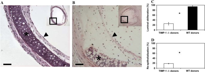
Obliterative bronchiolitis (OB) is a major cause of allograft dysfunction after lung transplantation and is thought to result from immunologically mediated airway epithelial destruction and luminal fibrosis. Matrix metalloproteinases (MMPs) and tissue inhibitors of metalloproteinases (TIMPs) have been implicated in the regulation of lung inflammation, airway epithelial repair, and extracellular matrix remodeling and therefore may participate in the pathogenesis of OB. The goals of this study were to determine the expression profiles of MMPs and TIMPs and the role of TIMP-1 in the development of airway obliteration using the murine heterotopic tracheal transplant model of OB. We demonstrate the selective induction of MMP-3, MMP-9, MMP-12, and TIMP-1 in a temporally restricted manner in tracheal allografts compared with isografts. In contrast, the expression of MMP-7, TIMP-2, and TIMP-3 was decreased in allografts relative to isografts during the period of graft rejection. TIMP-1 protein localized to epithelial, mesenchymal, and inflammatory cells in the tracheal grafts in a temporally and spatially restricted manner. Using TIMP-1-deficient mice, we demonstrate that the absence of TIMP-1 in the donor trachea or the allograft recipient reduced luminal obliteration and increased re-epithelialization in the allograft compared with wild-type control at 28 d after transplantation. Our findings provide direct evidence that TIMP-1 contributes to the development of airway fibrosis in the heterotopic tracheal transplant model, and suggest a potential role for this proteinase inhibitor in the pathogenesis of OB in patients with lung transplant.
Figures


Similar articles
-
Role of airway epithelial injury in murine orthotopic tracheal allograft rejection.Ann Thorac Surg. 2006 Oct;82(4):1226-33. doi: 10.1016/j.athoracsur.2006.03.122. Ann Thorac Surg. 2006. PMID: 16996912
-
FK506 combined with GM6001 prevents tracheal obliteration in a mouse model of heterotopic tracheal transplantation.Transpl Immunol. 2019 Dec;57:101244. doi: 10.1016/j.trim.2019.101244. Epub 2019 Sep 14. Transpl Immunol. 2019. PMID: 31526865
-
Increased matrix metalloproteinase-2 and membrane type 1 matrix metalloproteinase activity and expression in heterotopically transplanted murine tracheas.J Heart Lung Transplant. 2004 Feb;23(2):218-27. doi: 10.1016/S1053-2498(03)00112-8. J Heart Lung Transplant. 2004. PMID: 14761770
-
The heterotopic tracheal allograft as an animal model of obliterative bronchiolitis.Respir Res. 2001;2(3):169-83. doi: 10.1186/rr55. Epub 2001 Apr 5. Respir Res. 2001. PMID: 11686882 Free PMC article. Review.
-
Human and murine obliterative bronchiolitis in transplant.Proc Am Thorac Soc. 2007 Jan;4(1):37-43. doi: 10.1513/pats.200605-107JG. Proc Am Thorac Soc. 2007. PMID: 17202290 Free PMC article. Review.
Cited by
-
Matrix metalloproteinases in lung: multiple, multifarious, and multifaceted.Physiol Rev. 2007 Jan;87(1):69-98. doi: 10.1152/physrev.00022.2006. Physiol Rev. 2007. PMID: 17237343 Free PMC article. Review.
-
Interleukin-1β in tendon injury enhances reparative gene and protein expression in mesenchymal stem cells.Front Vet Sci. 2022 Aug 11;9:963759. doi: 10.3389/fvets.2022.963759. eCollection 2022. Front Vet Sci. 2022. PMID: 36032300 Free PMC article.
-
The effects of artocarpin on wound healing: in vitro and in vivo studies.Sci Rep. 2017 Nov 15;7(1):15599. doi: 10.1038/s41598-017-15876-7. Sci Rep. 2017. PMID: 29142215 Free PMC article.
-
Regression of allograft airway fibrosis: the role of MMP-dependent tissue remodeling in obliterative bronchiolitis after lung transplantation.Am J Pathol. 2011 Sep;179(3):1287-300. doi: 10.1016/j.ajpath.2011.05.032. Epub 2011 Jul 16. Am J Pathol. 2011. PMID: 21763265 Free PMC article.
-
Metalloproteinases and their inhibitors: regulators of wound healing.Int J Biochem Cell Biol. 2008;40(6-7):1334-47. doi: 10.1016/j.biocel.2007.10.024. Epub 2007 Oct 26. Int J Biochem Cell Biol. 2008. PMID: 18083622 Free PMC article. Review.
References
-
- Trulock EP, Edwards LB, Taylor DO, Boucek MM, Berkeley MK, Hertz MI. Registry of the International Society for Heart and Lung Transplantation: twenty-second official adult lung and heart-lung transplant report-2005. J Heart Lung Transplant 2005;24:956–967. - PubMed
-
- Estenne M, Hertz M. Bronchiolitis obliterans after human lung transplantation. Am J Respir Crit Care Med 2002;166:440–444. - PubMed
-
- Boehler A, Kesten S, Weder W, Speich R. Bronchiolitis obliterans after lung transplantation. Chest 1998;114:1411–1426. - PubMed
-
- Estenne M, Maurer JR, Boehler A, Egan JJ, Frost A, Hertz M, Mallory GB, Snell GI, Yousem S. Bronchiolitis obliterans syndrome 2001: an update of the diagnostic criteria. J Heart Lung Transplant 2001;21:297–310. - PubMed
-
- Neuringer IP, Aris RM, Burns KA, Bartolotta TL, Chalermskulrat W, Randell SH. Epithelial kinetics in mouse heterotopic tracheal allografts. Am J Transplant 2002;2:410–419. - PubMed
Publication types
MeSH terms
Substances
Grants and funding
LinkOut - more resources
Full Text Sources
Molecular Biology Databases
Research Materials
Miscellaneous
