. 2006 Jan 10;45(1):1-10.
doi: 10.1021/bi052130w.

Pentapeptide repeat proteins

Affiliations

Pentapeptide repeat proteins

Matthew W Vetting et al. Biochemistry. .

Abstract

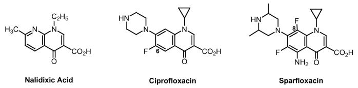
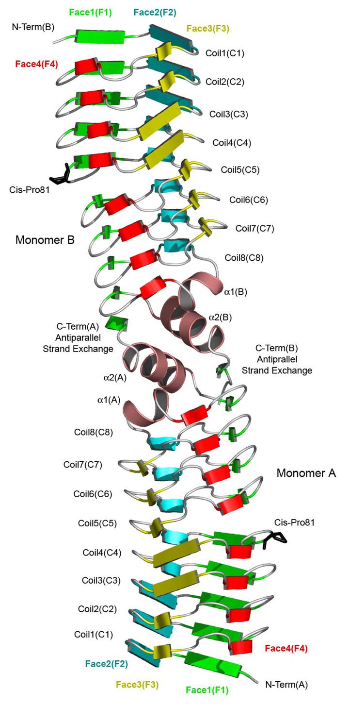
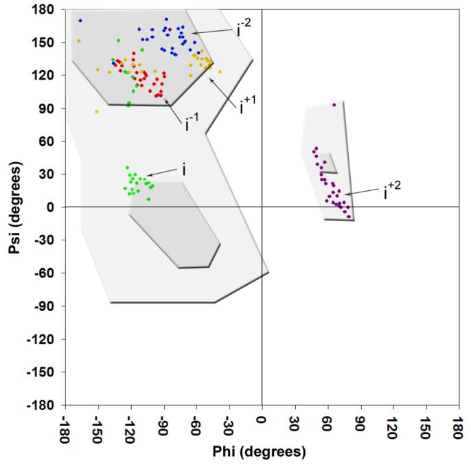
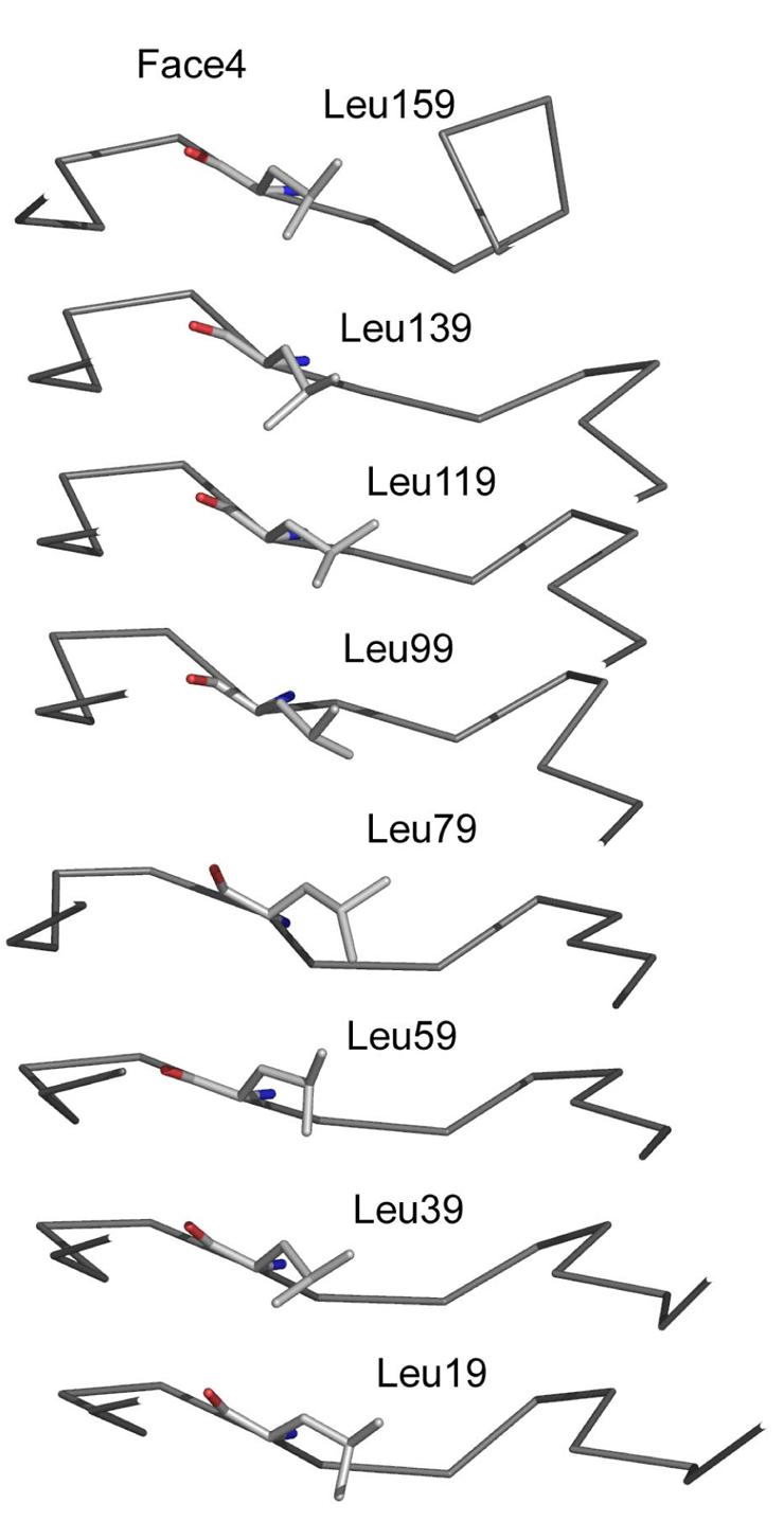
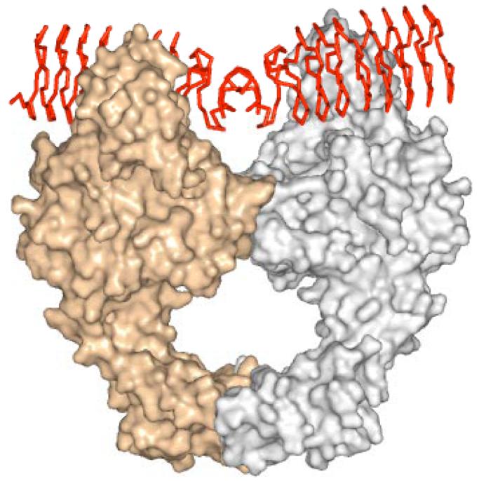
The pentapeptide repeat protein (PRP) family has more than 500 members in the prokaryotic and eukaryotic kingdoms. These proteins are composed of, or contain domains composed of, tandemly repeated amino acid sequences with a consensus sequence of [S,T,A,V][D,N][L,F][S,T,R][G]. The biochemical function of the vast majority of PRP family members is unknown. The three-dimensional structure of the first member of the PRP family was determined for the fluoroquinolone resistance protein (MfpA) from Mycobacterium tuberculosis. The structure revealed that the pentapeptide repeats encode the folding of a novel right-handed quadrilateral beta-helix. MfpA binds to DNA gyrase and inhibits its activity. The rod-shaped, dimeric protein exhibits remarkable similarity in size, shape, and electrostatics to DNA.


Figures

Scheme 1
Scheme 1
Figure 1
Figure 1
Ribbon diagram of the Mycobacterium tuberculosis MfpA dimer. The four faced of the quadrilateral β-helix are colored green (face 1), blue (face 2), yellow (face 3) and red (face 4).
Figure 2
Figure 2
Structure-based sequence alignment of MfpA. Regions comprising α1 and α2 are shaded salmon.
Figure 3
Figure 3
Electron density and molecular model of MfpA. Shown is 2Fo-Fc electron density contoured at 1σ and residues comprising the second coil.
Figure 4
Figure 4
Ramachandran plot of the first 165 residues that make up the right-handed quadrilateral β-helix domain of MfpA. Pentapeptide repeat positions are colored blue (i−2), red (i−1), green (i), yellow (i+1) and purple (i+2). The most favored and additionally allowed phi-psi orientations are shaded dark and light grey, respectively.
Figure 5
Figure 5
Side chain and main chain interactions of the pentapeptide repeat type II turns.
Figure 6
Figure 6
The interior phenylalanine stack on face 3 of MfpA.
Figure 7
Figure 7
The interior leucine stack on face 4 of MfpA
Figure 8
Figure 8
Model for the interaction of MfpA with DNA gyrase. DNA gyrase monomers are presented as silver and gold surface renderings, while the MfpA dimer is shown as a red Cα trace.
Figure 9
Figure 9
Genomic environment of M. tuberculosis MfpA. The four upstream genes colored in red are termed the “conservon” in other Actinomyces species.

References

    1. Black K, Buikema WJ, Haselkorn R. The hglK Gene is required for Localization of Heterocyst-Specific Glycolipids in the Cyanobacterium Anabena sp. Strain PCC 7120. J. Bact. 1995;177:6440–6448. - PMC - PubMed
    1. Bateman A, Murzin AG, Teichman SA. Structure and Distribution of Pentapeptide Repeats in Bacteria. Prot. Sci. 1998;7:1477–1480. - PMC - PubMed
    1. Altschul SF, Gish W, Miller W, Myers EW, Lipman DJ. Basic Alignment Search Tool. J. Mol. Biol. 1990;215:403–410. - PubMed
    1. Kolter R, Moreno F. Genetics of Ribosomally Synthesized Peptide Antibiotics. Ann. Rev. Microbiol. 1992;46:141–163. - PubMed
    1. Krogh A, Brown M, Mian IS, Sjolander K, Haussler D. Hidden Markov Models in Computational Biology. J. Mol. Biol. 1994;235:1501–1531. - PubMed

Publication types

MeSH terms