. 2006 Jan 10;45(1):167-74.
doi: 10.1021/bi051729t.

A new approach to producing functional G alpha subunits yields the activated and deactivated structures of G alpha(12/13) proteins

Affiliations

A new approach to producing functional G alpha subunits yields the activated and deactivated structures of G alpha(12/13) proteins

Barry Kreutz et al. Biochemistry. .

Abstract

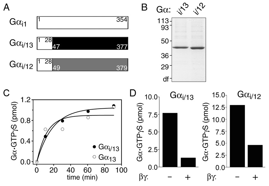
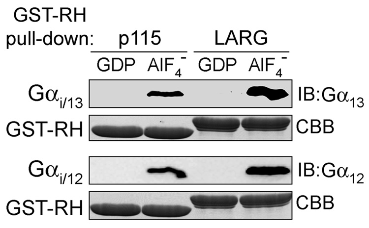
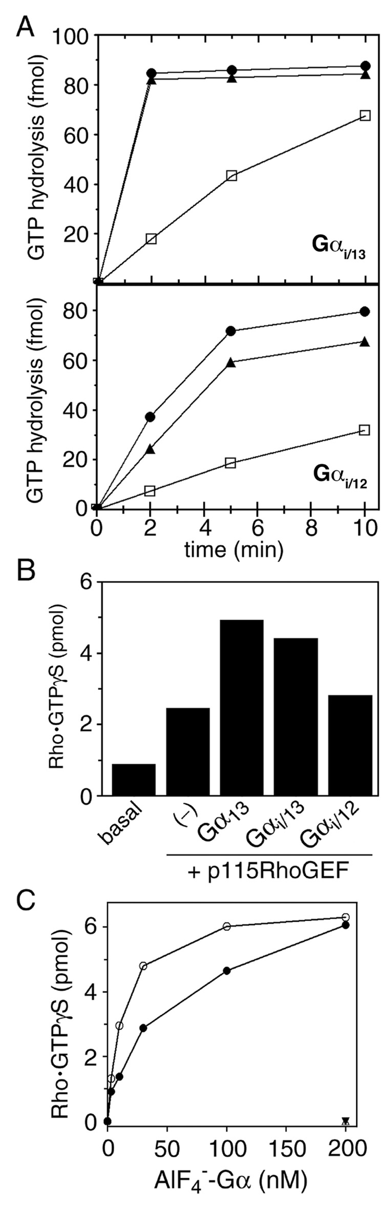
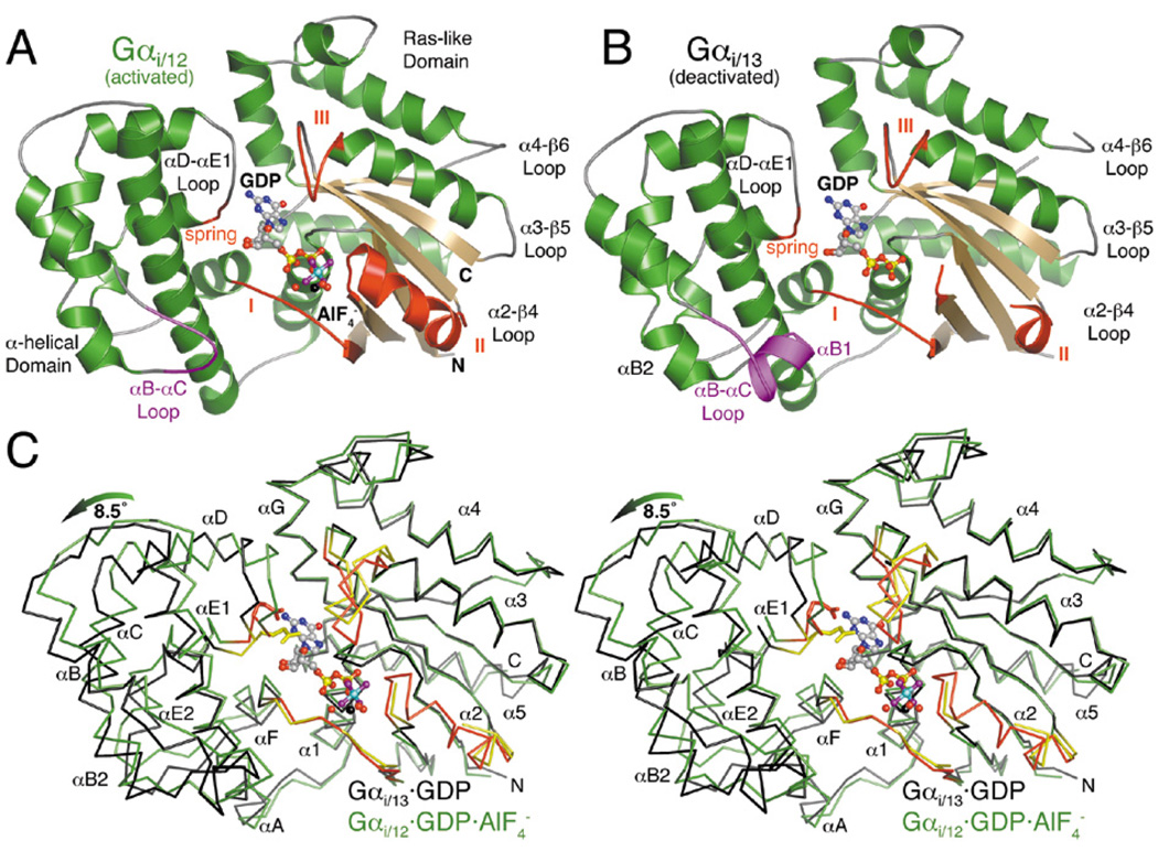
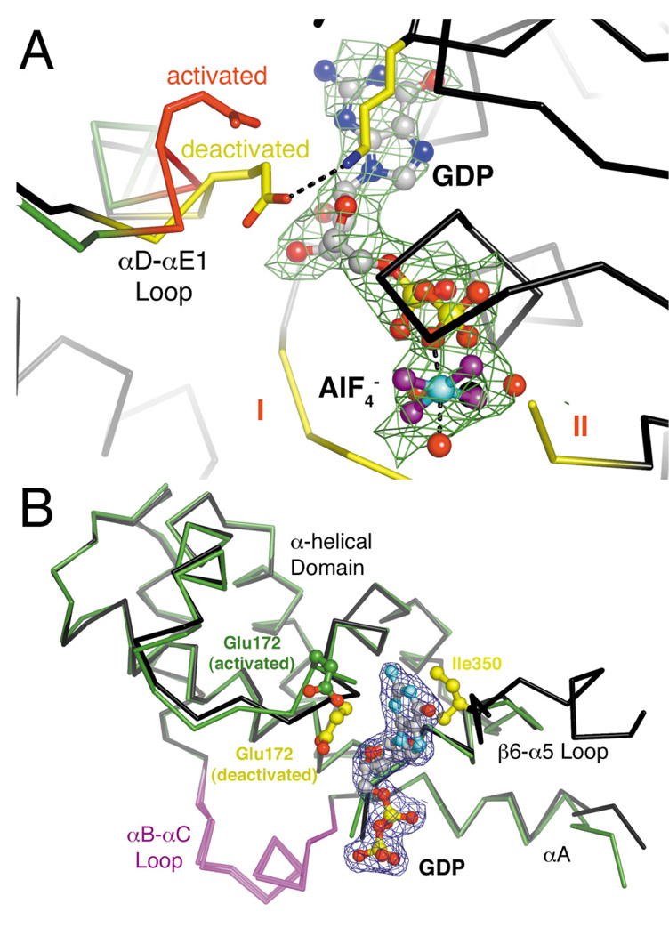
The oncogenic G(12/13) subfamily of heterotrimeric G proteins transduces extracellular signals that regulate the actin cytoskeleton, cell cycle progression, and gene transcription. Previously, structural analyses of fully functional G alpha(12/13) subunits have been hindered by insufficient amounts of homogeneous, functional protein. Herein, we report that substitution of the N-terminal helix of G alpha(i1) for the corresponding region of G alpha12 or G alpha13 generated soluble chimeric subunits (G alpha(i/12) and G alpha(i/13)) that could be purified in sufficient amounts for crystallographic studies. Each chimera bound guanine nucleotides, G betagamma subunits, and effector proteins and exhibited GAP responses to p115RhoGEF and leukemia-associated RhoGEF. Like their wild-type counterparts, G alpha(i/13), but not G alpha(i/12), stimulated the activity of p115RhoGEF. Crystal structures of the G alpha(i/12) x GDP x AlF4(-) and G alpha(i/13) x GDP complexes were determined using diffraction data extending to 2.9 and 2.0 A, respectively. These structures reveal not only the native structural features of G alpha12 and G alpha13 subunits, which are expected to be important for their interactions with GPCRs and effectors such as G alpha-regulated RhoGEFs, but also novel conformational changes that are likely coupled to GTP hydrolysis in the G alpha(12/13) class of heterotrimeric G proteins.


Figures

Figure 1
Figure 1
Generation of Gαi/13 and Gαi/12 chimeric proteins. (A) Schematic representation of the Gαi/13 and Gαi/12 chimera. Residues of Gαi1 are shown in white, and numbers within the colored regions of the Gαi/13 or Gαi/12 chimera correspond to original numbering of residues contributed by Gα13 (black) or Gα12 (gray). (B) Purified His6-Gαi/13 or His6-Gαi/12 (3 µg) were subjected to SDS-PAGE and stained with Coomassie Brilliant Blue. The position and apparent molecular weight of standard proteins (in kDa) is indicated. (C) GTPγS binding to Gαi/13 (●) or wild-type Gα13 (○) was measured at 30°C. Data are the mean of duplicate determinations. (D) Gαi/13 or Gαi/12 functionally interact with Gβγ. GTPγS binding to Gαi/13 or Gαi/12 (3 µg each) was measured either in the absence or presence of Gβγ (20 µg) as indicated. GTPγS binding was quantified after 90 min at 30°C. Data are the mean of duplicate determinations.
Figure 2
Figure 2
i/13 or Gαi/12 chimera interact with RH domains in an activation-dependent manner. GST-p115-RH or GST-LARG-RH were incubated with purified His6-Gαi/13 (upper panels) or His6-Gαi/12 (lower panels), either in the absence or presence of AlF4, and then GST-RH was pulled down using glutathione Sepharose 4B resin. Protein eluted from washed beads was resolved by SDS-PAGE and either immunoblotted (IB) with anti-Gα13 (B860) or anti-Gα12 (J168) antibody, or stained by Coomassie Brilliant Blue (CBB).
Figure 3
Figure 3
Activity of purified Gαi/13 and Gαi/12 in reconstitution assays. (A) Single-turnover hydrolysis of GTP bound to Gαi/13 (upper panel) or Gαi/12 (lower panel) was measured at 15°C in the absence (□) or presence of 100 nM GST-p115-RH (●) or 100 nM GST-LARG-RH (▲). Data are from one experiment, which is representative of three experiments. (B) Stimulation of p115RhoGEF activity by Gαi/13 but not Gαi/12. GTPγS binding to His6-RhoA was measured after incubation for 5 min at 30°C in the presence of p115RhoGEF (5 nM) and indicated AlF4-activated Gα subunit (100 nM). Data are the mean of duplicate determinations from one experiment, which is representative of three experiments. (C) Dose-dependent stimulation of p115RhoGEF activity by Gαi/13. GTPγS binding to His6-RhoA by p115RhoGEF (5 nM) was evaluated after 5 min at 30°C, and included the indicated concentration of either AlF4-activated wild-type Gα13 (○) or Gαi/13 (●). Samples lacking RhoA but containing p115RhoGEF and either AlF4-activated wild-type Gα13 (△) or Gαi/13 (▼) were assayed in parallel.
Figure 4
Figure 4
Structures of Gα12/13 subunits in activated and deactivated states. (A) The activated Gαi/12·GDP·AlF4 complex. The region of the αB-αC loop distinct from that of Gαi/13 is colored purple. The three conformationally flexible “switch regions” of the Gα subunit are red and labeled with Roman numerals (I–III). A fourth, apparently Gα12/13-specific element (the “spring”) is likewise colored red. The Mg2+·GDP·AlF4 ligand complex is shown as a ball-and-stick model, with carbons colored gray, oxygens red, nitrogens blue, phosphates yellow, fluorines purple, aluminum cyan and magnesium black. Waters are shown as red spheres. (B) The deactivated Gαi/13·GDP complex adopts an unusually open conformation in which the α-helical domain has rotated ~8.5° away from the Ras-like domain. Switch II is almost completely disordered, and switch III has rotated away (up in the figure) from the nucleotide binding site. (C) Stereo view of the Cα traces of Gαi/12·GDP·AlF4 (green with red switch regions) and Gαi/13·GDP (black with yellow switch regions), superimposed using their Ras-like domains. The glutamate side chain in the “spring” of each α-helical domain (E175 in Gα12, E172 in Gα13) is shown as a stick model.
Figure 5
Figure 5
The active sites and α-helical domains of activated and deactivated Gα12/13 subunits. (A) The activation-dependent “spring” of the α-helical domain. In the deactivated (GDP-bound) state, Gαi/13-E172 (yellow) is extended to form a salt bridge with Lys292 and packs against the nucleotide. In the activated (AlF4-bound) state, the spring (red) adopts a curled conformation and Gαi/12-Glu175 does not contact the bound nucleotide. Only the Ras-like domain of Gαi/13 is shown for clarity. Electron density from a 2.5 σ |Fo|-|Fc| omit map from the Gαi/12·GDP·AlF4 crystal structure is shown as green wire cage. (B) Superposition of the activated and deactivated conformations of the Gα13 α-helical domain. The Cα-trace of the α-helical domain (residues 74 to 201) and the β6-α5 loop (residues 345 to 357) of the Gαi/13·GDP structure are colored black, and the α-helical domain from the Gα13/i-5·p115RhoGEF complex is colored green. The αD-αE1 loop in the α-helical domain and the linker between the Ras-like and α-helical domains (N-terminal to αA) exhibit the most profound conformational changes upon nucleotide hydrolysis. Electron density from a 1.0 σ 2|Fo|-|Fc| map derived from the Gαi/13·GDP crystal structure is shown as green wire cage.

References

    1. Neves SR, Ram PT, Iyengar R. G protein pathways. Science. 2002;296:1636–1639. - PubMed
    1. Hollinger S, Hepler JR. Cellular regulation of RGS proteins: modulators and integrators of G protein signaling. Pharmacol Rev. 2002;54:527–559. - PubMed
    1. Berman DM, Kozasa T, Gilman AG. The GTPase-activating protein RGS4 stabilizes the transition state for nucleotide hydrolysis. J Biol Chem. 1996;271:27209–27212. - PubMed
    1. Tesmer JJG, Berman DM, Gilman AG, Sprang SR. Structure of RGS4 bound to AlF4−-activated Giα1: stabilization of the transition state for GTP hydrolysis. Cell. 1997;89:251–261. - PubMed
    1. Hepler JR, Gilman AG. G proteins. Trends Biochem Sci. 1992;17:383–387. - PubMed

Publication types

MeSH terms

Substances