. 2005 Dec;1(4):e45.
doi: 10.1371/journal.ppat.0010045. Epub 2005 Dec 30.

Bordetella pertussis, the causative agent of whooping cough, evolved from a distinct, human-associated lineage of B. bronchiseptica

Affiliations

Bordetella pertussis, the causative agent of whooping cough, evolved from a distinct, human-associated lineage of B. bronchiseptica

Dimitri A Diavatopoulos et al. PLoS Pathog. 2005 Dec.

Abstract

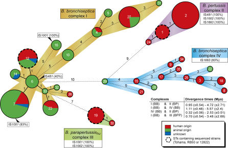
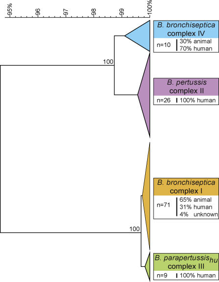
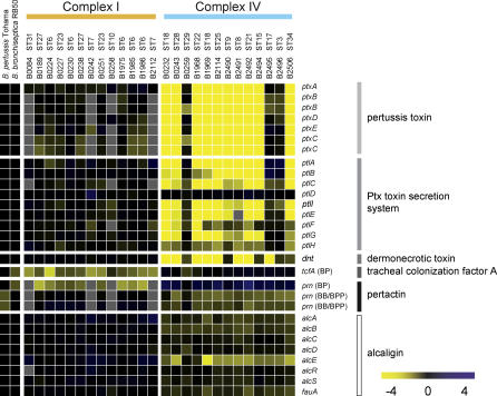
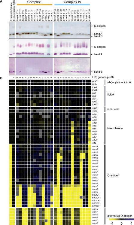
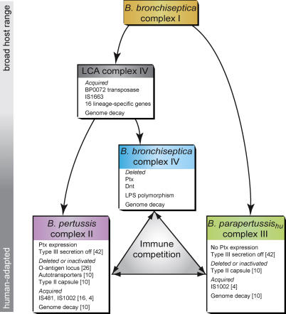
Bordetella pertussis, B. bronchiseptica, B. parapertussis(hu), and B. parapertussis(ov) are closely related respiratory pathogens that infect mammalian species. B. pertussis and B. parapertussis(hu) are exclusively human pathogens and cause whooping cough, or pertussis, a disease that has resurged despite vaccination. Although it most often infects animals, infrequently B. bronchiseptica is isolated from humans, and these infections are thought to be zoonotic. B. pertussis and B. parapertussis(hu) are assumed to have evolved from a B. bronchiseptica-like ancestor independently. To determine the phylogenetic relationships among these species, housekeeping and virulence genes were sequenced, comparative genomic hybridizations were performed using DNA microarrays, and the distribution of insertion sequence elements was determined, using a collection of 132 strains. This multifaceted approach distinguished four complexes, representing B. pertussis, B. parapertussis(hu), and two distinct B. bronchiseptica subpopulations, designated complexes I and IV. Of the two B. bronchiseptica complexes, complex IV was more closely related to B. pertussis. Of interest, while only 32% of the complex I strains were isolated from humans, 80% of the complex IV strains were human isolates. Comparative genomic hybridization analysis identified the absence of the pertussis toxin locus and dermonecrotic toxin gene, as well as a polymorphic lipopolysaccharide biosynthesis locus, as associated with adaptation of complex IV strains to the human host. Lipopolysaccharide structural diversity among these strains was confirmed by gel electrophoresis. Thus, complex IV strains may comprise a human-associated lineage of B. bronchiseptica from which B. pertussis evolved. These findings will facilitate the study of pathogen host-adaptation. Our results shed light on the origins of the disease pertussis and suggest that the association of B. pertussis with humans may be more ancient than previously assumed.


Conflict of interest statement

Competing interests. The authors have declared that no competing interests exist.

Figures

Figure 1
Figure 1. Minimum Spanning Tree of B. bronchiseptica, B. pertussis, and B. parapertussis
The tree was based on the sequence of seven housekeeping genes. Individual genes were split into five subloci, and a categorical clustering was performed. In the minimum spanning tree, sequence types sharing the highest number of single locus variants were connected first. Each circle represents a sequence type (ST) the size of which is related to the number of isolates within that particular ST. Colors within circles indicate host distribution. The numbers between connected STs represent the number of different subloci between those STs. The clonal complexes (I, II, III, and IV) are indicated by colored strips between connected STs. ST16 (B. bronchiseptica complex I) harbors the B. parapertussisov strains. STs containing strains of which the genome has been sequenced (B. pertussis Tohama, B. parapertussis 12822 or B. bronchiseptica RB50) are indicated by a thickset, dashed line. The distribution of the insertion sequence elements IS481, IS1001, IS1002, and IS1663 is shown in boxes (see also Table S1); numbers between parentheses indicate the percentage of strains that contained the ISE as determined by PCR amplification. The divergence times between B. bronchiseptica complexes I and IV and B. pertussis complex II are shown.
Figure 2
Figure 2. UPGMA Tree Based on the Analysis of the Pertactin Gene of Bordetella Isolates Used in the MLST Analysis
The DNA segment coding for the extracellular domain of pertactin (P.69) was used for analysis, with the exclusion of the repeat regions 1 and 2. Bootstrap values are shown for the nodes separating the complexes and are based on 500 bootstrap replicates. The scale indicates the genetic distance along the branches. Colors of the branches indicate the four complexes defined by MLST. The number of strains of each branch is shown in boxes, as well as the host distribution.
Figure 3
Figure 3. Gene Content of the Differentially Hybridizing Virulence Loci between B. bronchiseptica Complex I and IV, as Determined by CGH
Each column represents one strain. Strain numbers and STs are indicated above the columns. Each row represents one ORF (in B. bronchiseptica RB50 gene order), ORF designations are shown to the right of the rows. In the case of tcfA and prn, the origins of the probes are indicated between parentheses. The BP probe of tcfA was 100% similar to B. pertussis Tohama and 85.1% similar to B. bronchiseptica RB50. The BP prn probe was 100% similar to B. pertussis Tohama and 86% similar to B. parapertussis 12822 and B. bronchiseptica RB50. The BB/BPP prn probes were both 100% similar to B. parapertussis 12822 (BPP) and B. bronchiseptica RB50 (BB) and 86% similar to B. pertussis Tohama. The yellow-black-blue color scale indicates the hybridization value relative to the reference; references are B. bronchiseptica RB50, B. parapertussis 12822, and B. pertussis Tohama. For B. bronchiseptica RB50 and B. pertussis Tohama, the data in the figure are based on the genomic sequences. Yellow indicates decreased hybridization, black indicates hybridization values comparative to the references, and blue indicates gene duplications. Intermediate values indicate partial deletions or sequence divergence. Missing data are represented in gray.
Figure 4
Figure 4. Expression of LPS by B. bronchiseptica Complex I and Complex IV Strains and Gene Content Variation at the LPS Biosynthesis Locus
(A) Top: Electrophoretic LPS profiles obtained by tricine-SDS-PAGE and silver staining. Middle: Western blot of the same samples with mAb 36G3, which detects band A. Bottom: Western blot of the same samples with mAb BL8, which detects band B. (B) Gene content of the LPS biosynthesis locus as determined by CGH. See Figure 3 for details. For B. bronchiseptica RB50 and B. pertussis Tohama, the data in the figure are based on the genomic sequences. The genes wbmPQRSTU represent an alternative LPS O-antigen biosynthesis sublocus that is orthologous to the genes found in B. parapertussis 12822 [10] and B. bronchiseptica C7635E [26]. LPS genetic profiles as described in the text are indicated at the top of the columns. Color scale as in Figure 3. Missing data are represented in gray.
Figure 5
Figure 5. Model of the Evolution of the Mammalian Bordetellae
The bar on the left indicates increasing degrees of adaptation to the human host. Arrows indicate descent; double arrows between complexes indicate possible within-host immune competition. In boxes, genetic events are shown that may have played a role in speciation and niche adaptation. Numbers between parentheses refer to references. See text for details.

Similar articles

Cited by

References

    1. Gueirard P, Weber C, Le Coustumier A, Guiso N. Human Bordetella bronchiseptica infection related to contact with infected animals: Persistence of bacteria in host. J Clin Microbiol. 1995;33:2002–2006. - PMC - PubMed
    1. Bauwens JE, Spach DH, Schacker TW, Mustafa MM, Bowden RA. Bordetella bronchiseptica pneumonia and bacteremia following bone marrow transplantation. J Clin Microbiol. 1992;30:2474–2475. - PMC - PubMed
    1. Woolfrey BF, Moody JA. Human infections associated with Bordetella bronchiseptica . Clin Microbiol Rev. 1991;4:243–255. - PMC - PubMed
    1. van der Zee A, Groenendijk H, Peeters M, Mooi FR. The differentiation of Bordetella parapertussis and Bordetella bronchiseptica from humans and animals as determined by DNA polymorphism mediated by two different insertion sequence elements suggests their phylogenetic relationship. Int J Syst Bacteriol. 1996;46:640–647. - PubMed
    1. Mooi FR, van Loo IH, King AJ. Adaptation of Bordetella pertussis to vaccination: A cause for its reemergence? Emerg Infect Dis. 2001;7((3 Suppl)):526–528. - PMC - PubMed