Review
. 2005 Jul;2(3):396-409.
doi: 10.1602/neurorx.2.3.396.

Rodent models of focal stroke: size, mechanism, and purpose

Affiliations
Review

Rodent models of focal stroke: size, mechanism, and purpose

S Thomas Carmichael. NeuroRx. 2005 Jul.

Abstract

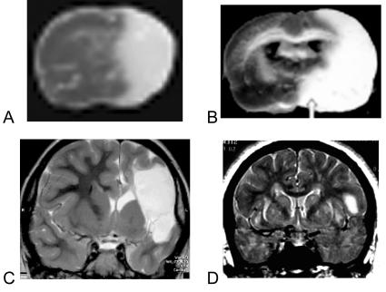
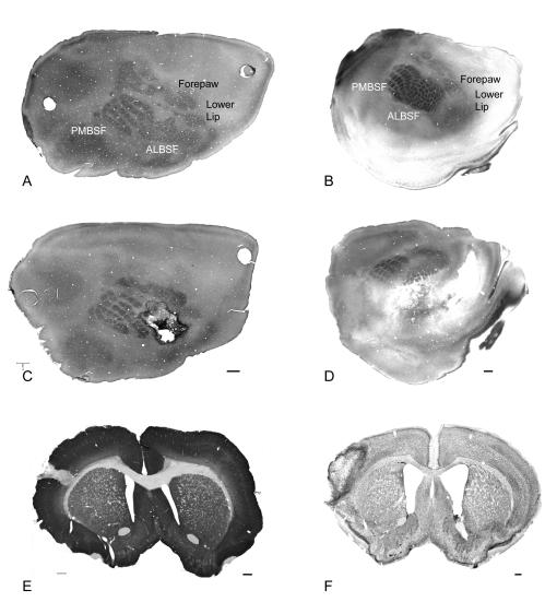
Rodent stroke models provide the experimental backbone for the in vivo determination of the mechanisms of cell death and neural repair, and for the initial testing of neuroprotective compounds. Less than 10 rodent models of focal stroke are routinely used in experimental study. These vary widely in their ability to model the human disease, and in their application to the study of cell death or neural repair. Many rodent focal stroke models produce large infarcts that more closely resemble malignant and fatal human infarction than the average sized human stroke. This review focuses on the mechanisms of ischemic damage in rat and mouse stroke models, the relative size of stroke generated in each model, and the purpose with which focal stroke models are applied to the study of ischemic cell death and to neural repair after stroke.


Figures

FIG. 1.
FIG. 1.
Stroke in rodent and human. A: TTC stained section from mouse with a 2-h suture occlusion of the MCA 1 day after the infarct was induced. The pale area is the infarcted tissue. B: TTC-stained section from rat with a permanent MCAO, one day after infarct was induced. C: T2-weighted MRI from a patient with a malignant infarction. This patient developed malignant edema and required hemicraniectomy. D: T2-weighted image from a patient with an infarct more representative of average human stroke size (Table 2). Panel A is reprinted with permission from Foerch et al. Serum S100B predicts a malignant course of infarction in patients with acute middle cerebral artery occlusion. Stroke 35:2160–2164. Copyright © 2004, Elsevier. All rights reserved. Panel B is reprinted with permission from Kim et al. Agmatine reduces infarct area in a mouse model of transient focal cerebral ischemia and protects cultured neurons from ischemia-like injury. Exp Neurol 189:122–130. Copyright © 2004, Lippincott, Williams and Wilkins. All rights reserved. Panel C is courtesy of Dr. Noriko Salamon, Division of Diagnostic Neuroradiology, Geffen School of Medicine at UCLA. Panel D is taken from the authors’ cases in the UCLA Stroke Service, Geffen School of Medicine at UCLA.
FIG. 2.
FIG. 2.
Focal stroke in rodent barrel field. Left column shows normal and post-stroke rat cortex; right column shows normal and post-stroke mouse cortex. A and C: Tangential sections of flattened rat cortex through cortical layer IV and stained for cytochrome oxidase and nestin shows the rat somatosensory body map. The barrel field (PMBSF and ALBSF) form a significant portion of the total surface area of the rat cortex. In panel C, a stroke has been produced in a portion of the barrel field and is highlighted by dark, nestin-positive reactive astrocytes. E: Coronal section through the infarct stained for MAP2 shows the relatively small total area occupied by the infarct. B and D: Tangential sections of mouse cortex through layer IV and stained for cytochrome oxidase show the somatosensory body map. The infarct is larger in the mouse, but also positioned within and adjacent to the barrel field. F: Coronal section through the infarct stained for Nissl. Bar in C = 1 mm and applied to A and C; bar in D = 400 μm and applied to B and D. Bar in E =1 mm; bar in F = 250 μm. PMBSF = posteromedial barrel field; ALBSF = anterolateral barrel field.

References

    1. American Heart Association, Heart Disease and Stroke Statistics Update 2004.http://www.americanheart.org/presenter.jhtml?identifier=3000090.
    1. Carmichael ST. Plasticity of cortical projections after stroke. Neuroscientist 9: 64–75, 2003. - PubMed
    1. Gladstone DJ, Black SE, Hakim AM. Heart and Stroke Foundation of Ontario Centre of Excellence in Stroke Recovery. Toward wisdom from failure: lessons from neuroprotective stroke trials and new therapeutic directions. Stroke 33: 2123–2236, 2003. - PubMed
    1. Cheng YD, Al-Khoury L, Zivin JA. Neuroprotection for ischemic stroke: two decades of success and failure. NeuroRx 1: 36–45, 2004. - PMC - PubMed
    1. Sugawara T, Fujimura M, Noshita N, Kim GW, Saito A, Hayashi T, et al. Neuronal death/survival signaling pathways in cerebral ischemia. NeuroRx 1: 17–25, 2004. - PMC - PubMed

Publication types