. 2006 Apr 15;395(2):319-29.
doi: 10.1042/BJ20051804.

Mode of action and subsite studies of the guluronan block-forming mannuronan C-5 epimerases AlgE1 and AlgE6

Affiliations

Mode of action and subsite studies of the guluronan block-forming mannuronan C-5 epimerases AlgE1 and AlgE6

Synnøve Holtan et al. Biochem J. .

Abstract

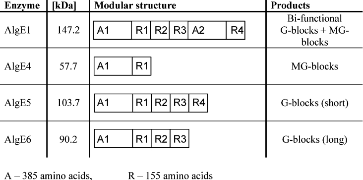
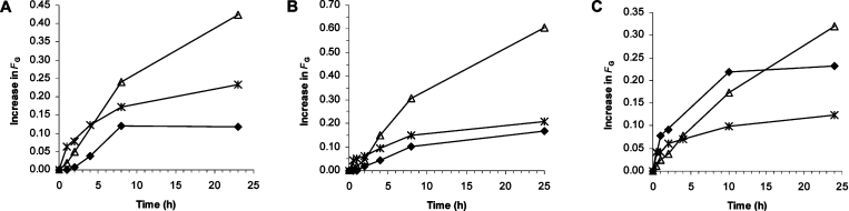
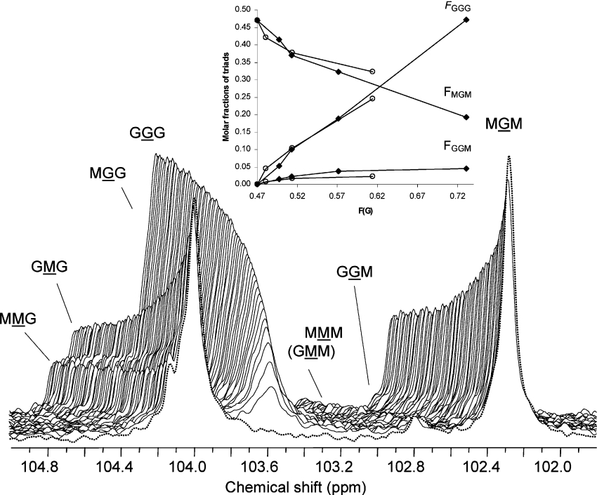
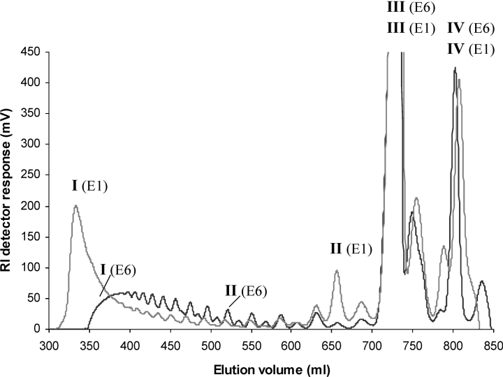
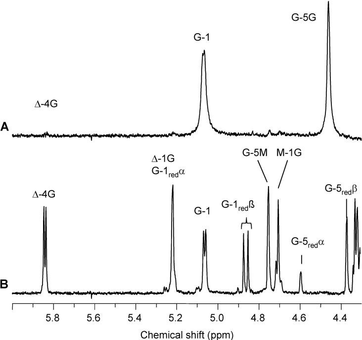
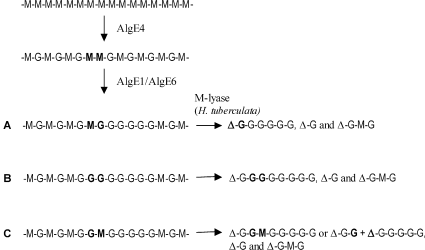
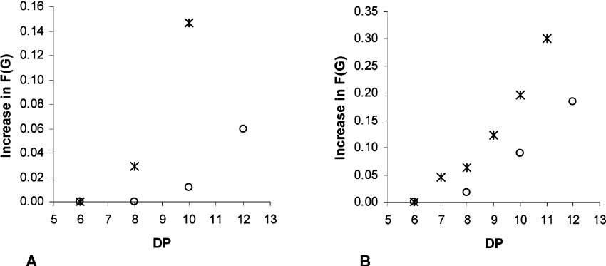
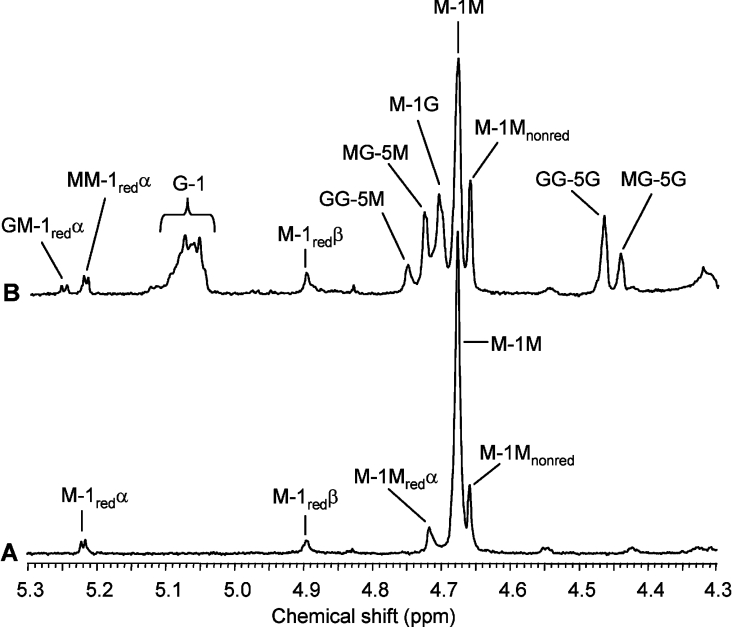
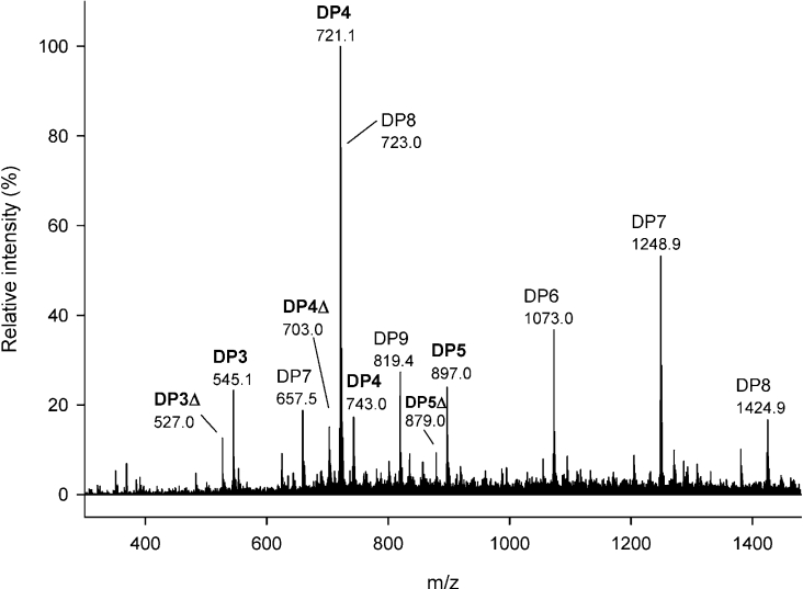
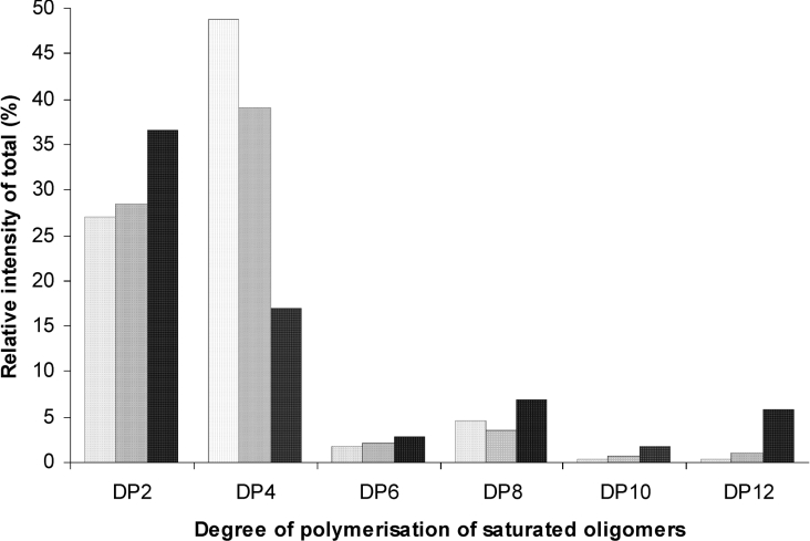
AlgE1, AlgE5 and AlgE6 are members of a family of mannuronan C-5 epimerases encoded by the bacterium Azotobacter vinelandii, and are active in the biosynthesis of alginate, where they catalyse the post-polymerization conversion of beta-D-mannuronic acid (M) residues into alpha-L-guluronic acid residues (G). All enzymes show preference for introducing G-residues neighbouring a pre-existing G. They also have the capacity to convert single M residues flanked by G, thus 'condensing' G-blocks to form almost homopolymeric guluronan. Analysis of the length and distribution of G-blocks based on specific enzyme degradation combined with size-exclusion chromatography, electrospray ionization MS, HPAEC-PAD (high-performance anion-exchange chromatography and pulsed amperometric detection), MALDI (matrix-assisted laser-desorption ionization)-MS and NMR revealed large differences in block length and distribution generated by AlgE1 and AlgE6, probably reflecting their different degree of processivity. When acting on polyMG as substrates, AlgE1 initially forms only long homopolymeric G-blocks >50, while AlgE6 gives shorter blocks with a broader block size distribution. Analyses of the AlgE1 and AlgE6 subsite specificities by the same methodology showed that a mannuronan octamer and heptamer respectively were the minimum substrate chain lengths needed to accommodate enzyme activities. The fourth M residue from the non-reducing end is epimerized first by both enzymes. When acting on MG-oligomers, AlgE1 needed a decamer while AlgE6 an octamer to accommodate activity. By performing FIA (flow injection analysis)-MS on the lyase digests of epimerized and standard MG-oligomers, the M residue in position 5 from the non-reducing end was preferentially attacked by both enzymes, creating an MGMGGG-sequence (underlined and boldface indicate the epimerized residue).


Figures

Figure 1
Figure 1. Molecular masses (kDa), modular protein structures and product specificities of four of the seven AlgE epimerases of A. vinelandii
Figure 2
Figure 2. Increase in FG as a function of epimerization time by AlgE1 (A), AlgE6 (B) and AlgE5 (C) on various substrates
Mannuronan (FM=1.0) (△), L. hyperborea leaf alginate (FG=0.49 and FGG=0.33) (*) and polyMG (FG=0.47 and FGG=0.0) (◆) as substrates.
Figure 3
Figure 3. 13C-NMR monitored AlgE6 epimerization of polyMG
A stacked plot of the anomeric region of 13C-NMR spectra (100 MHz) recorded during AlgE6 epimerization of 13C-1-enriched polyMG (FG=0.47 and FG=0.0) at 40 °C and p2H 7 of buffer solution (Mops) in 2H2O. The enzymatic reaction was carried out inside the NMR spectrometer. The final concentrations in 500 μl total volume in the NMR tube were 20 mg/ml polyMG-alginate, 5 mg/ml AlgE6 (as total protein) and 5 mM Ca2+. Molar ratio of enzyme/mannuronic acid residues susceptible to epimerization (FM=0.53) was 1:999. Spectra recorded after 19.9 min (FG=0.51) to 17.1 h reaction time (FG=0.77) with a time interval of 15.8 min between successive recordings are shown. Front spectrum (dotted line) is recorded before addition of epimerase. Inset: molar fractions of chain trisaccharide combinations versus degree of conversion (FG) for polyMG epimerized with the mannuronan C-5 epimerases AlgE1 (○) and AlgE6 (◆). Molar fractions were determined from the NMR spectra after 0, 4, 8 and 23 h reaction time for AlgE1, and after 0, 2, 4, 8 and 25 h reaction time for AlgE6. The epimerization reactions were then terminated. FGGM represents the molar fraction of G-block termination, i.e. the increase in number of G-blocks in the polymer chain during the epimerization reaction.
Figure 4
Figure 4. SEC of AlgE1- and AlgE6-epimerized polyMG (FG=0.55 for both enzymes) specific degraded by M-lyase from H. tuberculata
The oligo-uronic acids were chromatographed on three columns of Superdex 30 at a flow rate of 0.8 ml/min with 0.1 M ammonium acetate at room temperature. The eluent was monitored on-line with a refractive index detector. The DP and composition (sequence) of marked peaks [I−IV (E1) and (E6)] are given in Table 1. RI, refractive index.
Figure 5
Figure 5. 1H-NMR (400 MHz) spectra of the void (A) and major low-molecular-mass fraction (B) from SEC of the lyase digest of AlgE1-epimerized polyMG (FG=0.55)
The void and the low-molecular-mass fraction are denoted I (E1) and III (E1) respectively in the SEC chromatogram in Figure 4. Δ-4G and Δ-1G signals are produced upon lyase degradation. Δ denotes a 4-deoxy-L-erythro-hex-4-enepyranosyl uronate residue. Resonance signals from G residues (G-1red, G-5redα and G-5redβ) are dominating at the reducing end (B). The void fraction in (A) has FG≥0.97. The low-molecular-mass fraction has FG=0.5 and an alternating structure, determined as Δ-G-M-G with additional ESI-MS analysis confirming the molecular mass of DP4Δ (Table 1).
Figure 6
Figure 6. Illustration of the preferential enzyme attack by AlgE1 or AlgE6 of interspersed MM-sequences in polyMG (FG=0.47) and the following degradation products produced after treatment with M-lyase from H. tuberculata
(A, B) Most probable epimerization patterns achieving homogeneous G-blocks with an odd number of uronic acid residues (including the 4-deoxy-L-erythro-hex-4-enepyranosyl uronate residue), in addition to ΔG-M-G and Δ-G as main degradation products after lyase treatment of low epimerized polyMG-alginates. (C) Heterogeneous blocks with an odd number of residues, or shorter homogeneous G-blocks but with an even number of residues produced upon lyase degradation of epimerized material.
Figure 7
Figure 7. Minimum chain lengths (DP) of M- (*) and MG- (○) oligomers to accommodate activity of AlgE1 (A) and AlgE6 (B) epimerases
Oligomer samples were treated with the individual epimerases for 24 h, freeze-dried and analysed by 1H-NMR (300 and 500 MHz) spectroscopy in 2H2O at 90 °C. Initial values of FG were 0.0 and 0.47 for M- and MG-oligomers respectively.
Figure 8
Figure 8. 1H-NMR (500 MHz) spectra of control (non-epimerized) (A) and AlgE6-epimerized (B) mannuronate undecamer
The oligomannuronic acid sample in (A) was treated with the epimerase for 24 h and an increase in FG from 0.0 to 0.30 (FGG=0.15) (B). The α-anomeric resonance signal of the reducing end at 5.22 p.p.m. (A) shifts down-field to 5.24 p.p.m. when the penultimate residue is epimerized (B).
Figure 9
Figure 9. Negative ion ESI mass spectrum of mannuronate octamer after treatment with AlgE6 epimerase and G-lyase
Peak attributions: saturated tetramer, 721.1 [M−H+] and 743.0 [M+Na+−2H+]; saturated (unchanged) octamer, 723.0 [M+Na+−3H+/2]2− and 1424.9 [M−H+]; saturated pentamer 897.0 [M−H+]; saturated trimer, 545.1 [M−H+]; unsaturated tetramer, 703.0 [M−H+]; unsaturated trimer, 527.0 [M−H+]; unsaturated pentamer, 879.0 [M−H+]; saturated (unchanged) heptamer, 657.5 [M+3Na+–5H+/2]2− and 1248.9 [M−H+]; saturated (unchanged) hexamer, 1073.0 [M−H+]; and saturated (unchanged) nonamer, 819.4 [M+Na++NH4+−4H+/2]2−. Capillary voltage: 3.5 kV; solvent: aq. 50% methanol containing 1% ammonia.
Figure 10
Figure 10. Relative intensities of total of saturated oligomers in the M-lyase (H. tuberculata) digests of DP12 MG-oligomers
Control (non-epimerized), FG=0.47 (light-grey bars to the left); AlgE1-epimerized, FG=0.52 (dark-grey bars in the middle); AlgE6-epimerized, FG=0.65 (black bars to the right). The digests were analysed by FIA-MS in negative ion mode. Quantitative analysis was performed by integrating a ±10 p.p.m. window for each m/z and the contributions from the generated ions, [M−H+], [M−2H+]2−, [M−3H+]3−, [M−4H+]4−, were summed for each chain length. Only the chain lengths with an even number of residues are shown, due to the degradation pattern of the lyase and low intensities of odd numbered oligomers.

Similar articles

Cited by

References

    1. Painter T. J. Algal polysaccharides. In: Aspinall G. O., editor. The Polysaccharides, vol. 2. New York: Academic Press; 1983. pp. 195–285.
    1. Gorin P. A. J., Spencer J. F. T. Exocellular alginic acid from Azotobacter vinelandii. Can. J. Chem. 1966;44:993–998.
    1. Govan J. R. W., Fyfe J. A. M., Jarman T. R. Isolation of alginate-producing mutants of Pseudomonas fluorescens, Pseudomonas putida and Pseudomonas mendocina. J. Gen. Microbiol. 1981;125:217–220. - PubMed
    1. Ertesvåg H., Doseth B., Larsen B., Skjåk-Bræk G., Valla S. Cloning and expression of an Azotobacter vinelandii mannuronan C-5 epimerase gene. J. Bacteriol. 1994;176:2846–2853. - PMC - PubMed
    1. Ertesvåg H., Høidal H. K., Hals I. K., Rian A., Doseth B., Valla S. A family of modular type mannuronan C-5 epimerase genes controls alginate structure in Azotobacter vinelandii. Mol. Microbiol. 1995;16:719–731. - PubMed

Publication types

MeSH terms