Comparative Study
. 2006 Jan 23;203(1):189-201.
doi: 10.1084/jem.20050433. Epub 2006 Jan 3.

Role of mitochondrial glucocorticoid receptor in glucocorticoid-induced apoptosis

Affiliations
Comparative Study

Role of mitochondrial glucocorticoid receptor in glucocorticoid-induced apoptosis

Ronit Vogt Sionov et al. J Exp Med. .

Abstract

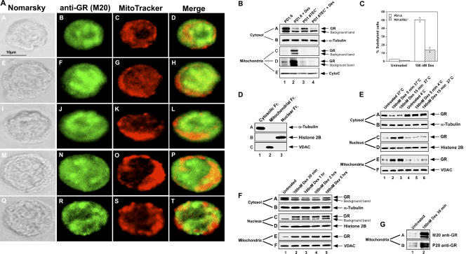
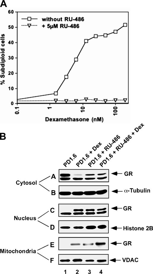
The mechanisms by which glucocorticoid receptor (GR) mediates glucocorticoid (GC)-induced apoptosis are unknown. We studied the role of mitochondrial GR in this process. Dexamethasone induces GR translocation to the mitochondria in GC-sensitive, but not in GC-resistant, T cell lines. In contrast, nuclear GR translocation occurs in all cell types. Thymic epithelial cells, which cause apoptosis of the PD1.6 T cell line in a GR-dependent manner, induce GR translocation to the mitochondria, but not to the nucleus, suggesting a role for mitochondrial GR in eliciting apoptosis. This hypothesis is corroborated by the finding that a GR variant exclusively expressed in the mitochondria elicits apoptosis of several cancer cell lines. A putative mitochondrial localization signal was defined to amino acids 558-580 of human GR, which lies within the NH2-terminal part of the ligand-binding domain. Altogether, our data show that mitochondrial and nuclear translocations of GR are differentially regulated, and that mitochondrial GR translocation correlates with susceptibility to GC-induced apoptosis.


Figures

Figure 1.
Figure 1.
(A) Sensitivity of various lymphoma and leukemia cell lines to Dex-induced apoptosis. Cells were incubated with 100 nM Dex for 20 h, and the DNA content measured by flow cytometry using propidium iodide. Percentage of subdiploid cells is given. (B) Dose–response to Dex. Cells were incubated with various concentrations of Dex and processed as in A. (C) Caspase 3 activation. Untreated or Dex-treated cells were stained for activated caspase 3 as described in Materials and methods. Percentage of positive cells is given. (D) Expression of mGR on lymphoma and leukemia cells. Untreated cells or cells treated with 100 nM Dex for 2 h were incubated with M20 antibodies to GR and FITC-conjugated goat anti–rabbit IgG (dashed line) or FITC-conjugated goat anti–rabbit IgG only (solid line). The fluorescence intensity was measured by flow cytometry.
Figure 2.
Figure 2.
(A) Intracellular staining of GR. PD1.6 cells were treated with 100 nM Dex for 2 h and the mitochondria were stained with 50 nM mitotracker. Rehydrated methanol-fixed cells were incubated with M20 antibodies to GR and FITC-conjugated goat anti–rabbit IgG, and visualized under confocal microscope. Five different cells are presented. (B) Dex induces mitochondrial translocation of GR in PD1.6, but not in PD1.6TEC cells. PD1.6 cells were incubated in the absence or presence of 100 nM Dex for 2 h before subcellular fractionation using the Oncogene fractionation kit. GR was detected by Western blotting using the PA1-511A antibody to GR (A, C, and D). (C and D) Data were obtained by two different exposure times. The blots were reprobed with antibodies to α-tubulin (B) and CytoC (D). CytoC could be used as a mitochondrial marker because the cells were harvested before any CytoC release. (C) Sensitivity of PD1.6 and PD1.6TEC cells to Dex-induced apoptosis. Cells were either untreated or treated with 100 nM Dex for 20 h, and percentage of apoptotic cells was determined as in Fig. 1 A. (D) The mitochondrial fraction is not contaminated by cytosolic or nuclear proteins. Subcellular fractions were run side by side on the same gel and analyzed by Western blotting using antibodies to α-tubulin (A), histone 2B (B), and VDAC (C), which react with cytosolic, nuclear, and mitochondrial fractions, respectively. (E) Dex induces a rapid, temperature-dependent translocation of GR to the nucleus and mitochondria in PD1.6 cells. PD1.6 cells were incubated in the absence or presence of 100 nM Dex for 5 and 15 min at 37°C (lanes 1–3) or 4°C (lanes 4–6) before subcellular fractionation as described in Materials and methods. GR was detected by Western blotting using the PA1-511A antibody to GR (A, C, and E). The blots were reprobed with antibodies to α-tubulin (B), histone H2B (D), and VDAC (F). (F) Dex induces sustained GR translocation to the nucleus and the mitochondria in PD1.6 cells. PD1.6 cells were incubated in the absence or presence of 100 nM Dex for 30 min to 5 h and processed as in E. (G) The mitochondrial GR is also immunoreactive to the M20 and P20 antibodies to GR. The mitochondrial samples 1 and 2 of F were rerun on gel and probed with either M20 or P20 antibodies.
Figure 3.
Figure 3.
(A–E) Dex induces GR translocation to the mitochondria in GC-sensitive PD1.6 cells (A), 2B4 cells (B), and thymocytes (C), but not in GC-resistant B10 (A), NB4 (D), S49 (E), and Jurkat (E) cells. Cells were incubated in the absence or presence of various Dex concentrations for 4 h before subcellular fractionation and Western blotting. In addition, NB4 cells were treated with 1 μM As2O3 (D, lanes 5 and 6), which is known to stabilize PML in these cells (reference 26).
Figure 4.
Figure 4.
(A) RU-486 prevents Dex-induced apoptosis of PD1.6 cells. PD1.6 cells were incubated with various concentrations of Dex in the absence or presence of 5 μM RU-486 for 20 h. Percentage of apoptosis was determined as in Fig. 1 A. (B) RU-486 induces mitochondrial translocation of GR in PD1.6 cells. Cells were incubated in the absence or presence of 5 μM RU-486 and/or 100 nM Dex for 2 h followed by subcellular fractionation and Western blotting.
Figure 5.
Figure 5.
TEC induces mitochondrial, but not nuclear, translocation of GR in TEC-sensitive PD1.6 cells. PD1.6, PD1.6TEC, and GR-deficient PD1.6Dex− cells were incubated alone or with TEC for 4 h. Nonadherent lymphoma cells were harvested and subjected to subcellular fractionation and Western blotting. The samples were harvested at an early stage after cocultivation with TEC, long before the onset of the apoptotic process. Thus, CytoC could be used as a marker for the mitochondria.
Figure 6.
Figure 6.
(A) MLS and NLS are located within different domains of GR. GR-negative 293 epithelial cells were transfected with plasmids encoding the indicated human GR variants. After 20 h, the cells were subjected to subcellular fractionation and GR was detected on Western blot using the PA1-511 antibody to GR. (B) α-Helix wheel model of the putative MLS located within aa 558–580 of human GR. The positive-charged arginine and lysine (red) and the hydrophilic threonine (orange) are located on the one side of the α-helix, whereas the hydrophobic aa leucine, isoleucine, valine, and tryptophane (blue) are located on the opposite side of the α-helix. Amino acids interacting with GC are labeled with gray numbers. This putative MLS is the α-Helix 3 of LBD. (C) The α-Helix 3 (aa 558–580) as it appears in the 1M2Z crystal structure. The same color labeling of aa is used as in B. (D) The MLS of GR resembles that of cytochrome C oxidase (COX). Amino acid sequence alignment between MLS of GR and MLS of COX. (E) The R564G and R575G mutants show reduced ability to enter the mitochondria. Mouse GFP-GR was point-mutated at aa 564 or 575 corresponding to the human R558 and R569, and the ability of these mutants to enter the mitochondria was determined as described in A. (F) The MLS (H3) of GR in the LBD crystal structure. The putative MLS is labeled in red in the 1M2Z crystal structure of a dimer complex of the human GR LBD (aa 521–777) bound to Dex and a Tif2 coactivator motif (reference 6).
Figure 7.
Figure 7.
(A and B) MLSCOX-GFP-GR is exclusively localized to the mitochondria. The mitochondria-directed GFP-GR variant was prepared by inserting the MLS of COX upstream to GFP-GR. HeLa cells (A), H1299 (B), or PC-3 (B) cells were transfected with either GFP-GR or MLSCOX-GFP-GR and stained with red mitrotracker to visualize the mitochondria before confocal microscopy. (C) MLSCOX-GFP-GR is more efficient in inducing apoptosis than GFP-GR. HeLa, L929 E8.2 A3, and PC-3 cells were transfected with plasmids encoding MLSCOX-GFP-GR or GFP-GR. After 48 h, the percentage of apoptotic GFP-positive cells (transfectants) was compared with that of GFP-negative cells (nontransfectants) of the same sample. (D) NLS-defective GRK513-515A induces apoptosis of HeLa cells. Cells were transfected with plasmids encoding GFP-GRwt, GFP-GRK513-515A, or pEGFP-F. After 48 h, the percentage of apoptotic cells was determined as in C. (E and F) A nucleus-directed GFP-GR variant (NLSTAg-GFP-GR) induces apoptosis of HeLa (E) and H1299 (F) cells. Cells were transfected with plasmids encoding GFP-GR, MLSCOX-GFP-GR, NLSTAg-GFP-GR, or pEGFP-F. After 48 h, the percentage of apoptotic cells was assessed as in C.

Similar articles

Cited by

References

    1. Haarman, E.G., G.J. Kaspers, and A.J. Veerman. 2003. Glucocorticoid resistance in childhood leukaemia: mechanisms and modulation. Br. J. Haematol. 120:919–929. - PubMed
    1. Tissing, W.J., J.P. Meijerink, M.L. den Boer, and R. Pieters. 2003. Molecular determinants of glucocorticoid sensitivity and resistance in acute lymphoblastic leukemia. Leukemia. 17:17–25. - PubMed
    1. Greenstein, S., K. Ghias, N.L. Krett, and S.T. Rosen. 2002. Mechanisms of glucocorticoid-mediated apoptosis in hematological malignancies. Clin. Cancer Res. 8:1681–1694. - PubMed
    1. Lu, N.Z., and J.A. Cidlowski. 2004. The origin and functions of multiple human glucocorticoid receptor isoforms. Ann. NY Acad. Sci. 1024:102–123. - PubMed
    1. Schaaf, M.J., and J.A. Cidlowski. 2002. Molecular mechanisms of glucocorticoid action and resistance. J. Steroid Biochem. Mol. Biol. 83:37–48. - PubMed

Publication types