. 2006 Jan 2;172(1):79-90.
doi: 10.1083/jcb.200508085.

Embryonic stem cells assume a primitive neural stem cell fate in the absence of extrinsic influences

Affiliations

Embryonic stem cells assume a primitive neural stem cell fate in the absence of extrinsic influences

Simon R Smukler et al. J Cell Biol. .

Abstract

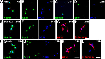
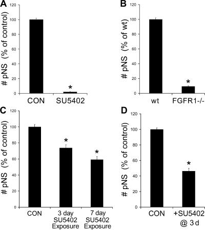
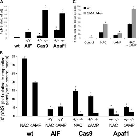
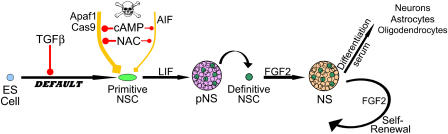
The mechanisms governing the emergence of the earliest mammalian neural cells during development remain incompletely characterized. A default mechanism has been suggested to underlie neural fate acquisition; however, an instructive process has also been proposed. We used mouse embryonic stem (ES) cells to explore the fundamental issue of how an uncommitted, pluripotent mammalian cell will self-organize in the absence of extrinsic signals and what cellular fate will result. To assess this default state, ES cells were placed in conditions that minimize external influences. Individual ES cells were found to rapidly transition directly into neural cells, a process shown to be independent of suggested instructive factors (e.g., fibroblast growth factors). Further, we provide evidence that the default neural identity is that of a primitive neural stem cell (NSC). The exiguous conditions used to reveal the default state were found to present primitive NSCs with a survival challenge (limiting their persistence and proliferation), which could be mitigated by survival factors or genetic interference with apoptosis.


Figures

Figure 1.
Figure 1.
ES cells rapidly transition into neural cells when placed in minimal conditions. (A and C) Immunocytochemical labeling showed that ES cells placed in minimal media conditions (for 4 h) initiated pronounced expression of the neural precursor markers nestin (A) and Sox1 (C). The nuclear stain DAPI was used to identify all cell nuclei within the field. (B and D) Undifferentiated ES cells (growing on a fibroblast feeder layer) exhibited Oct4 (B) but not nestin or Sox1 (D). The large Oct4 nuclei are feeder cells. (E) By 24 h, almost all cells expressed nestin and maintained some nuclear Oct4 expression and Sox1 (F). (G and H) Most of the ES-derived neural cells express NFM (G), and many express the early neuronal marker β3-tubulin (H; at 24 h). (I) Expression of nestin and Sox1 was confirmed by RT-PCR analysis of ES-derived neural cells (at 24 h). (J) Brachyury and GATA-1 (mesodermal markers), as well as HNF3β, HNF4, and GATA-4 (endodermal markers), were detected by RT-PCR in undifferentiated ES cells, though they were rapidly down-regulated within 24 h in minimal conditions. (K and L) After 3 d, most surviving ES-derived neural cells retain a neural precursor identity, maintaining expression of nestin (K) and Sox1 (L). (M and N) Differentiating cells with more elaborate morphologies were also apparent as β3-tubulin+ neurons (M) and O4+ oligodendrocytes (N). Bars: (A–D and K–N) 25 μm; (E–H) 10 μm.
Figure 2.
Figure 2.
The ES cell neural transition occurs by default without requirement for instructive factors. (A and B) Initiation of nestin (A) and Sox1 (B) expression was observed by immunocytochemical labeling within 4 h when ES cells were placed in PBS alone. (C and D) After 24 h in PBS, the few remaining viable cells expressed nestin (C) and Sox1 (D). The fragmented nuclei (punctuate DAPI staining) represent dead cells. (E–H) Pharmacological inhibition of FGF signaling using SU5402 (5 μM) did not prevent the rapid acquisition of neural markers by ES cells placed in minimal conditions for 24 h, with expression of nestin (E), Sox1 (F), NFM (G), and β3-tubulin (H) observed. (I–L) Similarly, ES cells harboring deletion of the fgfr1 gene displayed the typically observed neural markers by 24 h, expressing nestin (I), Sox1 (J), NFM (K), and β3-tubulin (L). Bars, 25 μm.
Figure 3.
Figure 3.
Primitive NSCs emerge from the ES cell default neural pathway. (A) When LIF was included in the minimal conditions, a small number of ES-derived neural precursor cells proliferated over 7 d to form clonally derived floating sphere colonies, termed primitive NSs. The primitive NS–initiating cell was termed a primitive NSC. (B) The primitive NS cells were found to coexpress nestin and Sox1 by immunocytochemical labeling, demonstrating their neural precursor identity. (C) RT-PCR analysis confirmed expression of nestin and Sox1 within primitive NSs (pNS). (D and E) When differentiated, primitive NSs yielded β3-tubulin+ neurons, GFAP+ astrocytes, and O4+ oligodendrocytes (D), whereas most cells retained a neural precursor phenotype, maintaining expression of nestin and Sox1 (E). Bars: (A) 100 μm; (B–D) 50 μm.
Figure 4.
Figure 4.
Primitive NSC proliferation is dependent on autogenously produced FGF signaling. (A) Pharmacological inhibition of FGF signaling throughout the primitive NS assay using SU5402 (5 μM) virtually abolished primitive NS formation (pNS). *, P < 0.001 versus control. (B) ES cells with a deletion of the fgfr1 gene displayed drastically reduced primitive NS production. *, P < 0.001 versus wild type (wt). (C) When SU5402 was included for 3 or 7 d of the primitive NS assay and then removed, the primitive NSCs resumed proliferation, and a substantial recovery of primitive NS formation was observed over an additional 7 d. *, P < 0.001 versus condition with maintained SU5402 (not depicted). (D) When SU5402 was added on day 3 of the primitive NS assay, proliferation of already proliferating primitive NSCs was impaired and fewer primitive NSs ensued. *, P < 0.001 versus control.
Figure 5.
Figure 5.
A survival challenge attributable to the minimal culture conditions limits the number of ES-derived neural precursors that survive to form primitive NSs. Extensive cell mortality was observed upon culture in the minimal conditions used for the default state assessment and primitive NS (pNS) assay. (A) Improving cell viability by inclusion of the survival factors NAC and the membrane-permeable cAMP analogue pCPT-cAMP (cAMP) dose-dependently enhanced primitive NS formation. *, P < 0.01 versus control. (B) Concurrent inclusion of 1 mM NAC and 100 μM pCPT-cAMP (a concentration at which peak effects were observed in A) in the primitive NS assay showed that they produced an additive, if not synergistic, effect. *, P < 0.001 versus control; , P < 0.001 versus control and versus each factor alone. (C) The effectiveness of NAC (1 mM) and pCPT-cAMP (100 μM) in promoting NS formation decreased dramatically with passaging of NSs. *, P < 0.001 versus respective survival factor primary spheres.
Figure 6.
Figure 6.
Genetic interference with apoptotic pathways enhances primitive NS generation and TGFβ inhibition independently cooperates with survival factors to promote primitive NS production. (A) ES cell lines with mutations in apoptotic pathway components aif, cas9, and apaf1 displayed considerably enhanced primitive NS (pNS) production compared with wild-type (wt) ES cells. *, P < 0.05 versus wt; , P < 0.001 versus wt and versus respective +/− genotype. (B) The primitive NS–promoting ability of NAC and pCPT-cAMP (cAMP) was assessed for each genotype of the apoptosis mutant ES cell lines. The effects of the survival factors were much reduced, though still considerable, in the aif mutant. Strikingly, the effects of NAC and pCPT-cAMP were drastically reduced in the cas9/− and apaf1/− ES cells. *, P < 0.001 versus wt in same survival factor, , P < 0.001 versus wt in same survival factor and versus respective +/− genotype in same survival factor. (C) The smad4/− ES cell line exhibited enhanced primitive NS production compared with wt ES cells. This independent enhancement was observed even in the presence of the survival factors NAC and pCPT-cAMP (cAMP), included individually or concurrently. *, P < 0.05 versus wt in same conditions; , P < 0.001 versus wt in same conditions and versus smad4/− in each factor alone.
Figure 7.
Figure 7.
ES cell–based model describing the ontogeny of NSCs. When placed in minimal conditions, free of inhibitory influences, ES cells will acquire a primitive NSC identity through a default mechanism. This transition is negatively regulated by TGFβ signaling. The nascent primitive NSC then undergoes a separate survival challenge in these minimal conditions, with apoptosis mediated primarily by the Apaf1–Cas9 pathway as well as contribution by the AIF apoptosis pathway. Survival can be promoted by NAC and cAMP, which mediate their effects largely through abrogation of the Apaf1–Cas9 pathway. In the presence of LIF, the primitive NSC remains in an undifferentiated, proliferative state and generates a primitive NS containing a subpopulation of more mature (FGF2 dependent) definitive NSCs. Upon dissociation of the primitive NS, the definitive NSCs will proliferate in an FGF2-dependent manner to generate an NS. In the presence of FGF2, this self-renewal passaging can be performed repeatedly. With the addition of serum, the NS can be differentiated to produce neurons, astrocytes, and oligodendrocytes.

References

    1. Bainter, J.J., A. Boos, and K.L. Kroll. 2001. Neural induction takes a transcriptional twist. Dev. Dyn. 222:315–327. - PubMed
    1. Baker, J.C., R.S. Beddington, and R.M. Harland. 1999. Wnt signaling in Xenopus embryos inhibits bmp4 expression and activates neural development. Genes Dev. 13:3149–3159. - PMC - PubMed
    1. Beddington, R.S., and E.J. Robertson. 1989. An assessment of the developmental potential of embryonic stem cells in the midgestation mouse embryo. Development. 105:733–737. - PubMed
    1. Bok, J., X.M. Zha, Y.S. Cho, and S.H. Green. 2003. An extranuclear locus of cAMP-dependent protein kinase action is necessary and sufficient for promotion of spiral ganglion neuronal survival by cAMP. J. Neurosci. 23:777–787. - PMC - PubMed
    1. Cai, J., J. Yang, and D.P. Jones. 1998. Mitochondrial control of apoptosis: the role of cytochrome c. Biochim. Biophys. Acta. 1366:139–149. - PubMed

Publication types

Substances