. 2006 Jan 2;172(1):103-13.
doi: 10.1083/jcb.200508001.

Distinct roles for Pax7 and Pax3 in adult regenerative myogenesis

Affiliations

Distinct roles for Pax7 and Pax3 in adult regenerative myogenesis

Shihuan Kuang et al. J Cell Biol. .

Abstract

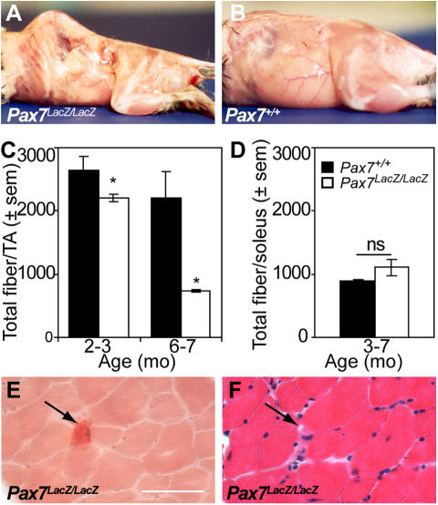
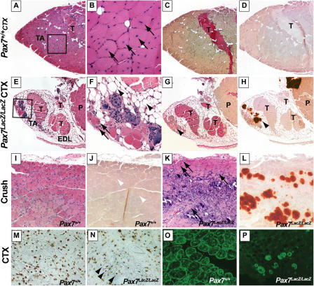
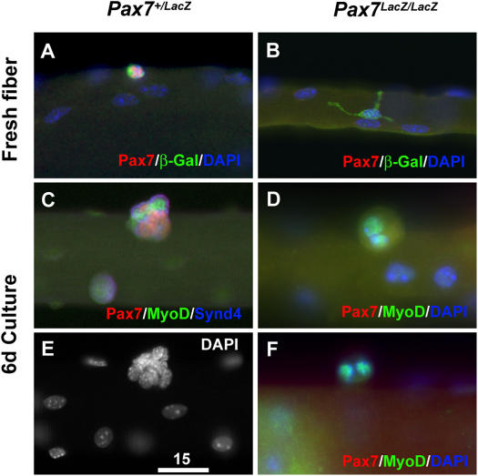
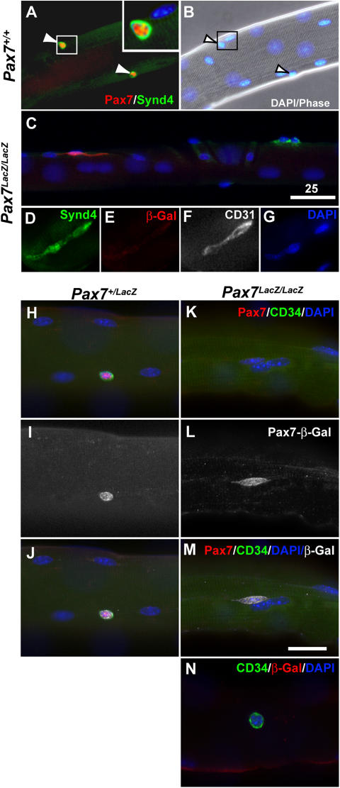
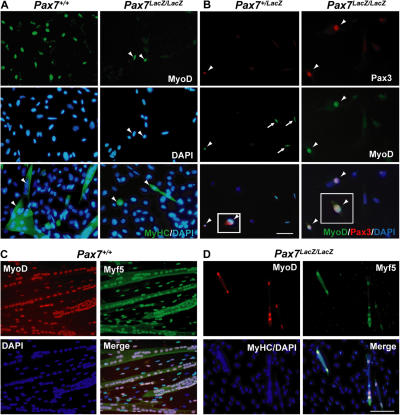
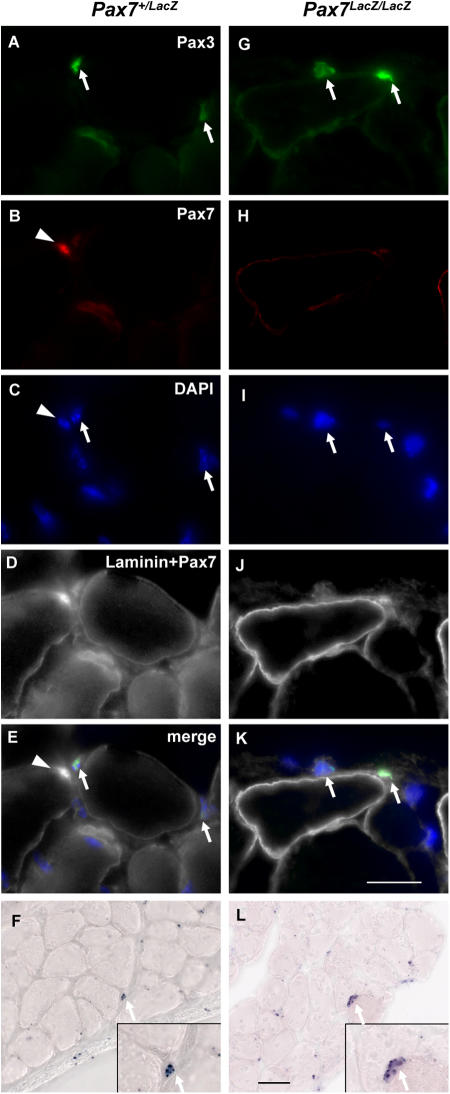
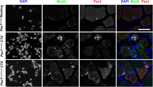
We assessed viable Pax7(-/-) mice in 129Sv/J background and observed reduced growth and marked muscle wasting together with a complete absence of functional satellite cells. Acute injury resulted in an extreme deficit in muscle regeneration. However, a small number of regenerated myofibers were detected, suggesting the presence of residual myogenic cells in Pax7-deficient muscle. Rare Pax3(+)/MyoD+ myoblasts were recovered from Pax7(-/-) muscle homogenates and cultures of myofiber bundles but not from single myofibers free of interstitial tissues. Finally, we identified Pax3+ cells in the muscle interstitial environment and demonstrated that they coexpressed MyoD during regeneration. Sublaminar satellite cells in hind limb muscle did not express detectable levels of Pax3 protein or messenger RNA. Therefore, we conclude that interstitial Pax3+ cells represent a novel myogenic population that is distinct from the sublaminar satellite cell lineage and that Pax7 is essential for the formation of functional myogenic progenitors from sublaminar satellite cells.


Figures

Figure 1.
Figure 1.
Accelerated muscle wasting in adult Pax7 lacZ/lacZ mice. (A and B) By 6 mo of age, Pax7 lacZ/lacZ (A) mice develop a curvature of the spine characteristic of extensive muscle wasting compared with wild-type littermates (B). (C) Total fiber number per TA is significantly reduced in Pax7 lacZ/lacZ mice compared with wild-type littermates (n = 4 and 5 for 2–4 mo and n = 2 and 2 for 6–7 mo for Pax7 + / + and Pax7 lacZ/lacZ, respectively). *, P < 0.05. (D) Total fiber number per soleus is not affected in Pax7 lacZ/lacZ mice compared with wild-type littermates (n = 2 for each genotype). (E and F) Small calcium deposits (arrows) stained with Alizarin red (E) are present in between Pax7 lacZ/lacZ myofibers, which otherwise appear normal, albeit smaller, on a spaced serial cross section stained with hematoxylin–eosin (F).
Figure 2.
Figure 2.
Profound regeneration deficit in Pax7 lacZ/l acZ muscle after acute injury. Lower hind limb sections from Pax7 + / + (A–D) or Pax7 lacZ/lacZ (E–H) mice 1 mo after CTX stained with hematoxylin–eosin (A, B, E, and F), Van Gieson's (C and G; pink stain), and Alizarin red (D and H; red stain). (B and F) Higher magnifications of A and E, respectively. Note the efficient muscle regeneration in Pax7 + / + TA characterized by numerous large centrally nucleated regenerating myofibers (B, arrows), with no extensive calcium deposition, adipogenesis, or fibrogenesis (A–D). In contrast, injured Pax7 lacZ/lacZ TA (E–H) displays only rare and small centrally nucleated myofibers (F, arrows); instead, the muscle was replaced by calcium deposits (H, arrowhead), adipocytes (F, arrowheads), or fibrosis (G, arrowhead). Sections from Pax7 + / + (I and J) and Pax7 lacZ/lacZ littermates (K and L) 10 d after crush injury reveal an efficient muscle regeneration process in Pax7 + / + TA (I), whereas only rare centrally nucleated myofibers are observed in Pax7 lacZ/lacZ TA (K, arrows). Instead, muscle is infiltrated by inflammatory cells and replaced by fibrosis and large calcium deposits (L). The arrowheads in J indicate normal regenerative fibers in wild type. Central myonuclei in regenerated TA myofibers from Pax7 + / + (M) and Pax7 lacZ/lacZ (N) muscle 10 d after CTX injection are BrdU+ (DAB, brown; N, arrowheads) after multiple BrdU injections during the period of regeneration. Low efficiency of muscle regeneration in the Pax7 lacZ/lacZ (P) is contrasted with the robust regeneration in Pax7 + / + (O), as demonstrated by the expression of Desmin (FITC, green). P, plantarus; T, tendon.
Figure 3.
Figure 3.
Remnant nonfunctional β-gal + cells associated with muscle fibers in Pax7 mutant mice. Expression of β-gal, knocked into the first coding exon of Pax7 gene to generate the Pax7 knockouts, in Pax7 + /lacZ (A) and Pax7 lacZ/lacZ (B) muscle fibers isolated from 1-mo-old mice. In Pax7 + /lacZ muscle fibers, expression of β-gal is always colocalized with Pax7 expression. In Pax7 lacZ/lacZ fibers, the β-gal+ cells display large cell bodies and extensive filopodia-like processes (B), which are in contrast to the smaller, round morphologies of the β-gal+ cells in the Pax7 + /lacZ fibers (A). (C–F) After 6 d of suspended culture of single muscle fibers from Pax7 + /lacZ mice, the Pax7+ cells proliferate and differentiate to form aggregates of cells that express Pax7, MyoD, or both (C and E). Under identical culture conditions, the β-gal+ cells associated with muscle fibers of Pax7 lacZ/lacZ mice are unable to proliferate or differentiate (D and F). Bar, 15 μm.
Figure 4.
Figure 4.
Absence of satellite cell marker expression associated with adult Pax7 lacZ/lac Z muscle fibers. (A and B) Pax7 and Syndecan4 are coexpressed in satellite cells on freshly isolated Pax7 + / + EDL fibers. Arrowheads show satellite cells and corresponding nuclei stained with DAPI. (C) In the Pax7 lacZ/lacZ myofibers, few β-gal+ cells (red) were identified, but they did not express the satellite cell marker Syndecan4 (green). (C–G) Conversely, the low frequencies of Syndecan4+ cells associated with Pax7 lacZ/lacZ myofibers were negative for β-gal but positive for the endothelial cells marker CD31. (H–J) Similarly, Pax7+ and β-gal+ cells on the Pax7 + /lacZ myofibers are also positive for another satellite cell marker, CD34. (K–N) In the Pax7 lacZ/lacZ myofibers, however, rare β-gal+ and CD34+ cells were never colocalized: the β-gal+ cells were negative for CD34, and the CD34+ cells were negative for β-gal. Bars, 25 μm.
Figure 5.
Figure 5.
Identification of Pax7-independent myogenic cells expressing Pax3. (A, top and middle) Cell suspensions from Pax7 + / + hind limb muscles grown in growth media for 15 h yielded large numbers of MyoD+ cells (FITC) compared with rare MyoD+ cells (arrowheads) observed in Pax7 lacZ/lacZ preparations. (bottom) Myogenic cells obtained from both Pax7 + / + and Pax7 lacZ/lacZ hind limb muscles were capable of terminal differentiation as shown by MyHC expression (green; arrowheads) and fusion following 3 d in differentiation media. (B) Double immunostaining for Pax3 (red) and MyoD (green) of cell suspensions from Pax7 + /lacZ or Pax7 lacZ/lacZ limb muscle revealed that Pax3 and MyoD were coexpressed in certain myogenic cells (arrowheads). Cells expressing MyoD but not Pax3 were also detected in Pax7 + / + and Pax7 lacZ/lacZ cultures (arrows; not depicted). Insets show enlargements of the cells to the left. (C and D) Myogenic cells derived from cultures myofiber bundles of Pax7 + / + (C) and Pax7 lacZ/lacZ (D) mice. Myofiber bundle (20–30 fibers) were plated in a Matrigel-coated dish and culture in growth medium for 3 d followed by 1 d in differentiation medium. The green signals in the merged panels are intensified to help visualize myotubes. Bars, 12.5 μm.
Figure 6.
Figure 6.
Pax3 expression in Pax7 lacZ/lac Z and Pax7 +/lacZ musculature. (A–E and G–K) Triple staining of adult EDL muscle sections with laminin, Pax7, and Pax3 antibodies plus DAPI labeling of nuclei. Pax3+ cells (arrows), located in the interstitial space outside the basal lamina of myofibers, exist in similar frequency in both Pax7 + /lacZ (C) and Pax7 lacZ/lacZ (D) muscles. Note that the Pax7+ cell (arrowheads) in the Pax7 + /lacZ is located in a sublaminar position. Bar, 10 μm. (F and L) Pax3 in situ hybridization on Pax7 + /lacZ (F) and Pax7 lacZ/lacZ (L) diaphragm also revealed the presence of Pax3+ cells in both genotypes. Bar, 25 μm.
Figure 7.
Figure 7.
Myogenic specification of Pax3 + cells during muscle regeneration. In resting muscle, Pax3+ cells did not coexpress MyoD. In regenerating Pax7 lacZ/lacZ and Pax7 + /lacZ muscles 48 h after CTX injection, many Pax3+ cells coexpressed MyoD, indicating that these cells had acquired a myogenic fate. Note that in Pax7 + /lacZ, the majority of MyoD+ cells did not express Pax3 and are likely expressing Pax7. Bar, 20 μm.

Similar articles

Cited by

References

    1. Asakura, A., P. Seale, A. Girgis-Gabardo, and M.A. Rudnicki. 2002. Myogenic specification of side population cells in skeletal muscle. J. Cell Biol. 159:123–134. - PMC - PubMed
    1. Beauchamp, J.R., L. Heslop, D.S. Yu, S. Tajbakhsh, R.G. Kelly, A. Wernig, M.E. Buckingham, T.A. Partridge, and P.S. Zammit. 2000. Expression of CD34 and Myf5 defines the majority of quiescent adult skeletal muscle satellite cells. J. Cell Biol. 151:1221–1234. - PMC - PubMed
    1. Ben-Yair, R., and C. Kalcheim. 2005. Lineage analysis of the avian dermomyotome sheet reveals the existence of single cells with both dermal and muscle progenitor fates. Development. 132:689–701. - PubMed
    1. Bittner, R.E., C. Schofer, K. Weipoltshammer, S. Ivanova, B. Streubel, E. Hauser, M. Freilinger, H. Hoger, A. Elbe-Burger, and F. Wachtler. 1999. Recruitment of bone-marrow-derived cells by skeletal and cardiac muscle in adult dystrophic mdx mice. Anat. Embryol. (Berl.). 199:391–396. - PubMed
    1. Blau, H.M., T.R. Brazelton, and J.M. Weimann. 2001. The evolving concept review of a stem cell: entity or function? Cell. 105:829–841. - PubMed

Publication types

MeSH terms