. 2006 Jan 2;172(1):151-62.
doi: 10.1083/jcb.200506184.

Endothelial FAK is essential for vascular network stability, cell survival, and lamellipodial formation

Affiliations

Endothelial FAK is essential for vascular network stability, cell survival, and lamellipodial formation

Rickmer Braren et al. J Cell Biol. .

Abstract

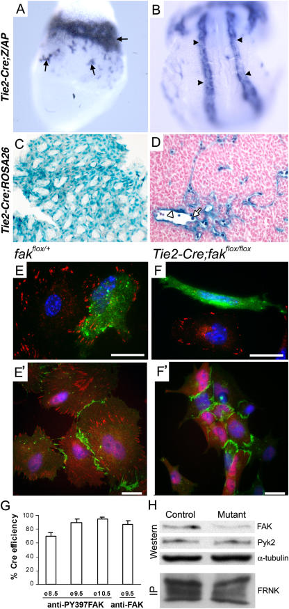
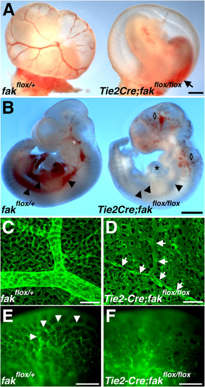
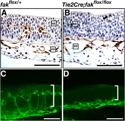
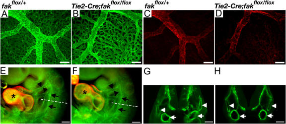
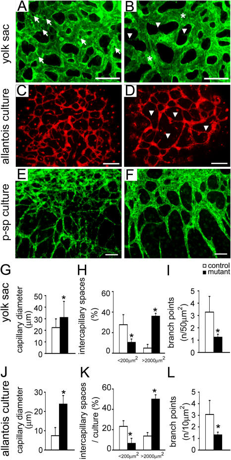
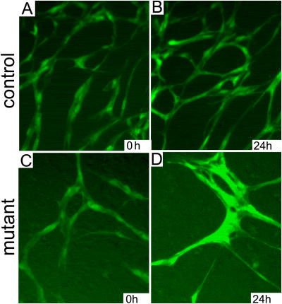
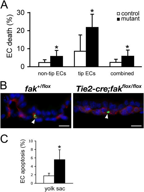
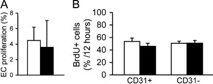
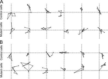
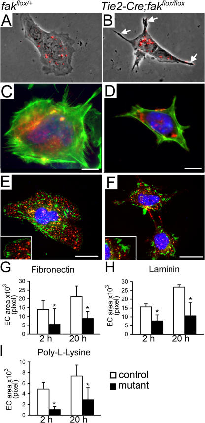
Morphogenesis of a vascular network requires dynamic vessel growth and regression. To investigate the cellular mechanism underlying this process, we deleted focal adhesion kinase (FAK), a key signaling mediator, in endothelial cells (ECs) using Tie2-Cre mice. Targeted FAK depletion occurred efficiently early in development, where mutants exhibited a distinctive and irregular vasculature, resulting in hemorrhage and lethality between embryonic day (e) 10.5 and 11.5. Capillaries and intercapillary spaces in yolk sacs were dilated before any other detectable abnormalities at e9.5, and explants demonstrate that the defects resulted from the loss of FAK and not from organ failure. Time-lapse microscopy monitoring EC behavior during vascular formation in explants revealed no apparent decrease in proliferation or migration but revealed increases in cell retraction and death leading to reduced vessel growth and increased vessel regression. Consistent with this phenotype, ECs derived from mutant embryos exhibited aberrant lamellipodial extensions, altered actin cytoskeleton, and nonpolarized cell movement. This study reveals that FAK is crucial for vascular morphogenesis and the regulation of EC survival and morphology.


Figures

Figure 1.
Figure 1.
Cre efficiently mediates gene excision early in the vasculature of Tie2-C re embryos. (A and B) AP-stained embryos at e7.5 (A) and e8.5 (B) show Cre activity in blood islands (arrows) and dorsal aortae (arrowheads). (C) A LacZ-stained YS at e11.5. (D) Paraffin section of e11.5 LacZ-stained embryo shows Cre activity in ECs (arrowhead) and blood cells (arrow). (E and F) FAK expression in cells from e9.5 embryos stained with anti-CD31 (green), DAPI (blue), and anti-Y397pFAK (red, E and F) or anti-FAK (red, E' and F'). Bars, 10 μm. (G) Cre-mediated FAK depletion was measured by the percentage of FAK CD31+ cells in mutants at e8.5, 9.5, and 10.5 relative to controls. Each represents the mean ± SEM (error bars) of 100 CD31+ cells in three replicate (for anti-Y397FAK) and five replicate (for anti-FAK) experiments. (H) Expression of FAK, Pyk2, and FRNK by Western or IP analyses of e9.5 embryos.
Figure 2.
Figure 2.
Tie2-Cre–mediated FAK depletion causes hemorrhaging and vascular defects at e10.5. (A and B) Images of freshly dissected YSs (A) and embryos (B). Arrow, bleeding; diamonds, dilated sinusoidal vessels; asterisk, heart; arrowheads, major vessels. Bars, 1 mm. (C and D) Microangiographs of CD31-stained YSs. Note that only the remnants of large vessels are identifiable in the mutant (D, arrows). (E and F) Microangiographs of CD31-stained heads. Arrowheads, internal carotid artery. Note the abnormally wide irregular vessels in the mutant. Bars, 200 μm.
Figure 3.
Figure 3.
Impaired sprouting angiogenesis into the NE in e10.5 mutants. (A and B) Paraffin cross sections of the apical head stained for CD31 (brown). Note multiple small perineural plexus (PP) capillaries in the NE in the control (A) but not in the mutant NE (B). (C and D) 100-μm cross sections of the heads show the capillary network in the control NE (C) but not in the mutant (D). Brackets, width of the NE. Bars, 100 μm.
Figure 4.
Figure 4.
e9.5 mutants display no gross defects in major vascular structures. (A–D) Microangiographs of e9.5 YSs stained with Abs to CD31 (green, A and B) and SMαA (red, C and D). (E–H) Microangiographs of whole-mount (E and F) and thick-sectioned (G and H) embryos labeled with Abs to CD31 and SMαA. Arrows, dorsal aortae; black arrowheads, aortic arches; asterisks, heart chambers; white arrowheads, paired cardinal veins. Bars, 200 μm.
Figure 5.
Figure 5.
Dilated capillaries and intercapillary spaces are primary defects. (A and B) Microangiographs of e9.5 control (A) and mutant (B) YSs. Note fewer small intercapillary spaces (arrows), widened capillaries (asterisk), and more incomplete sprouts (arrowheads) in the mutant. Bars, 100 μm. (C and D) Microangiographs of control (C) and mutant (D) allantoic explants. Arrowheads, widened intercapillary spaces. Bars, 200 μm. (E and F) Microangiographs of control (E) and mutant (F) P-Sp explants. Note a reduction in network complexity and wider vessels in the mutants. (G–L) Quantification of capillary diameters, intercapillary space sizes, and number of branch points in YSs (G–I) and allantoic explants (J–L). Error bars represent SEM. *, P < 0.05.
Figure 6.
Figure 6.
Still images of capillaries in P-Sp explants. Control (A) and mutant (C) explants show comparable capillary networks at 0 h, but at 24 h, there were fewer, wider vessels in the mutant (D) compared with the control (B).
Figure 7.
Figure 7.
Increased death in FAK-deficient ECs. (A) EC death in P-Sp explants as detected by time-lapse microscopy. (B) Cross sections of immunofluorescent stained e9.5 YSs. Red, CD31; green, TUNEL; arrowheads, TUNEL+ ECs. Bars, 10 μm. (C) Apoptotic rate of ECs in the YSs. Bar graph represents the mean ± SEM (error bars) from three independent experiments. A total of 10,175 control and 7,878 mutant ECs were counted. *, P < 0.05. Control, white bars; mutant, black bars.
Figure 8.
Figure 8.
Insignificant decrease in proliferation in FAK-deficient ECs. (A) EC proliferation in P-Sp explants as measured by time-lapse microscopy. (B) EC proliferation measured by in vivo BrdU labeling. Each bar represents the mean ± SEM (error bars) of 100 cells in three independent experiments. Control, white bars; mutant, black bars.
Figure 9.
Figure 9.
Migration paths of individual FAK-deficient ECs in P-Sp explants. Each point indicates the position (XY coordinates) of the cell at 30- (A, capillary ECs) or 28- (B, single ECs) min intervals. Positions of 10 consecutive intervals are shown. Each axis tick represents a distance of 10 μm.
Figure 10.
Figure 10.
Mutant ECs display defective cell morphology and locomotion. (A and B) Micrographs of ECs overlaid with DiI-Ac-LDL labeling (red). Mutant ECs display thin and spiky cell protrusions (arrows). (C–F) Immunofluorescent micrographs of actin organization (C and D) and focal adhesions (E and F) in ECs; insets, focal adhesions. Red, CD31; green, actin (C and D) or paxillin (E and F); blue, DAPI. Bars, 10 μm. (G–I) Time course of EC spreading on FN, LM, and PLL at 2 and 20 h. Each bar represents the ± SEM (error bars) of 50 cells examined in three replicate experiments. *, P < 0.05.

Similar articles

Cited by

References

    1. Abu-Ghazaleh, R., J. Kabir, H. Jia, M. Lobo, and I. Zachary. 2001. Src mediates stimulation by vascular endothelial growth factor of the phosphorylation of focal adhesion kinase at tyrosine 861, and migration and anti-apoptosis in endothelial cells. Biochem. J. 360:255–264. - PMC - PubMed
    1. Adini, I., I. Rabinovitz, J.F. Sun, G.C. Prendergast, and L.E. Benjamin. 2003. RhoB controls Akt trafficking and stage-specific survival of endothelial cells during vascular development. Genes Dev. 17:2721–2732. - PMC - PubMed
    1. Alavi, A., J.D. Hood, R. Frausto, D.G. Stupack, and D.A. Cheresh. 2003. Role of Raf in vascular protection from distinct apoptotic stimuli. Science. 301:94–96. - PubMed
    1. Ausprunk, D.H., and J. Folkman. 1977. Migration and proliferation of endothelial cells in preformed and newly formed blood vessels during tumor angiogenesis. Microvasc. Res. 14:53–65. - PubMed
    1. Beggs, H.E., D. Schahin-Reed, K. Zang, S. Goebbels, K.A. Nave, J. Gorski, K.R. Jones, D. Sretavan, and L.F. Reichardt. 2003. FAK deficiency in cells contributing to the basal lamina results in cortical abnormalities resembling congenital muscular dystrophies. Neuron. 40:501–514. - PMC - PubMed

Publication types

MeSH terms