

Monocyte-derived CXCL7 peptides in the marrow microenvironment
- PMID: 16391012
- PMCID: PMC1895768
- DOI: 10.1182/blood-2005-10-4285
Monocyte-derived CXCL7 peptides in the marrow microenvironment
Abstract
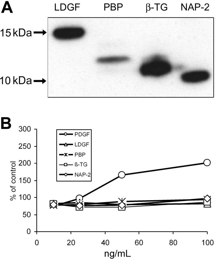
The marrow microenvironment consists of several different interacting cell types, including hematopoietic-derived monocyte/macrophages and nonhematopoietic-derived stromal cells. Gene-expression profiles of stromal cells and monocytes cultured together differ from those of each population alone. Here, we report that CXCL7 gene expression, previously described as limited to the megakaryocyte lineage, is expressed by monocytes cocultured with stromal cells. CXCL7 gene expression was confirmed by quantitative reverse transcriptase-polymerase chain reaction (RT-PCR), and secretion of protein was detected by enzyme-linked immunosorbent assay (ELISA) and Western blot. At least 2 stromal-derived activities, one yet to be identified, were required for optimal expression of CXCL7 by monocytes. NAP-2, the shortest form of CXCL7 detected in the coculture media, was confirmed to decrease the size and number of CFU-Meg colonies. The propeptide LDGF, previously reported to be mitogenic for fibroblasts, was not secreted by stimulated monocytes. The recombinant form of LDGF produced in a prokaryotic expression system did not have biologic activity in our hands. The monocytic source of CXCL7 was also detected by immunohistochemistry in normal bone marrow biopsies, indicating an in vivo function. We conclude that stromal-stimulated monocytes can serve as an additional source for CXCL7 peptides in the microenvironment and may contribute to the local regulation of megakaryocytopoiesis.
Figures


Similar articles
-
Functionally and phenotypically distinct subpopulations of marrow stromal cells are fibroblast in origin and induce different fates in peripheral blood monocytes.Stem Cells Dev. 2014 Apr 1;23(7):729-40. doi: 10.1089/scd.2013.0300. Epub 2013 Nov 23. Stem Cells Dev. 2014. PMID: 24131213 Free PMC article.
-
Human marrow stromal cells activate monocytes to secrete osteopontin, which down-regulates Notch1 gene expression in CD34+ cells.Blood. 2004 Jun 15;103(12):4496-502. doi: 10.1182/blood-2004-01-0256. Epub 2004 Mar 2. Blood. 2004. PMID: 14996707
-
Monocytes secrete CXCL7 to promote breast cancer progression.Cell Death Dis. 2021 Nov 17;12(12):1090. doi: 10.1038/s41419-021-04231-4. Cell Death Dis. 2021. PMID: 34789744 Free PMC article.
-
Regulation of production of activin A in human marrow stromal cells and monocytes.Exp Hematol. 1992 Nov;20(10):1235-42. Exp Hematol. 1992. PMID: 1426103
-
Canine CXCL7 and its functional expression in dendritic cells undergoing maturation.Vet Immunol Immunopathol. 2010 May 15;135(1-2):128-136. doi: 10.1016/j.vetimm.2009.11.011. Epub 2009 Nov 29. Vet Immunol Immunopathol. 2010. PMID: 20022386
Cited by
-
Specific molecular signatures predict decitabine response in chronic myelomonocytic leukemia.J Clin Invest. 2015 May;125(5):1857-72. doi: 10.1172/JCI78752. Epub 2015 Mar 30. J Clin Invest. 2015. PMID: 25822018 Free PMC article. Clinical Trial.
-
Proteome-based biomarkers in pancreatic cancer.World J Gastroenterol. 2011 Nov 28;17(44):4845-52. doi: 10.3748/wjg.v17.i44.4845. World J Gastroenterol. 2011. PMID: 22171124 Free PMC article. Review.
-
The role of the marrow microenvironment in hematopoietic stem cell transplantation.Cell Ther Transplant. 2010 Apr 1;2(7):7-12. doi: 10.3205/ctt-2009-en-000072.01. Cell Ther Transplant. 2010. PMID: 21243120 Free PMC article.
-
The role of platelet factor 4 in radiation-induced thrombocytopenia.Int J Radiat Oncol Biol Phys. 2011 Aug 1;80(5):1533-40. doi: 10.1016/j.ijrobp.2011.03.039. Int J Radiat Oncol Biol Phys. 2011. PMID: 21740995 Free PMC article.
-
The possible diagnostic and prognostic use of systemic chemokine profiles in clinical medicine—the experience in acute myeloid leukemia from disease development and diagnosis via conventional chemotherapy to allogeneic stem cell transplantation.Toxins (Basel). 2013 Feb 18;5(2):336-62. doi: 10.3390/toxins5020336. Toxins (Basel). 2013. PMID: 23430540 Free PMC article. Review.
References
-
- Dexter TM, Moore MAS. In vitro duplication and `cure' of haemopoietic defects in genetically anaemic mice. Nature. 1977;269: 412-414. - PubMed
-
- Novotny JR, Duehrsen U, Welch K, Layton JE, Cebon JS, Boyd AW. Cloned stromal cell lines derived from human Whitlock/Witte-type long-term bone marrow cultures. Exp Hematol. 1990;18: 775-784. - PubMed
-
- Slack JL, Nemunaitis J, Andrews DF, Singer JW. Regulation of cytokine and growth factor gene expression in human bone marrow stromal cells transformed with simian virus 40. Blood. 1990;75: 2319-2327. - PubMed
-
- Thalmeier K, Meissner P, Reisbach G, Falk M, Brechtel A, Dormer P. Establishment of two permanent human bone marrow stromal cell lines with long-term post irradiation feeder capacity. Blood. 1994;83: 1799-1807. - PubMed
-
- Roecklein BA, Torok-Storb B. Functionally distinct human marrow stromal cell lines immortalized by transduction with the human papilloma virus E6/E7 genes. Blood. 1995;85: 997-1005. - PubMed
Publication types
MeSH terms
Substances
Grants and funding
LinkOut - more resources
Full Text Sources
Other Literature Sources
Molecular Biology Databases
Research Materials
