. 2006 May 1;107(9):3555-63.
doi: 10.1182/blood-2005-10-4168. Epub 2006 Jan 3.

Enhancement of thrombogenesis by plasma fibronectin cross-linked to fibrin and assembled in platelet thrombi

Affiliations

Enhancement of thrombogenesis by plasma fibronectin cross-linked to fibrin and assembled in platelet thrombi

Jaehyung Cho et al. Blood. .

Abstract

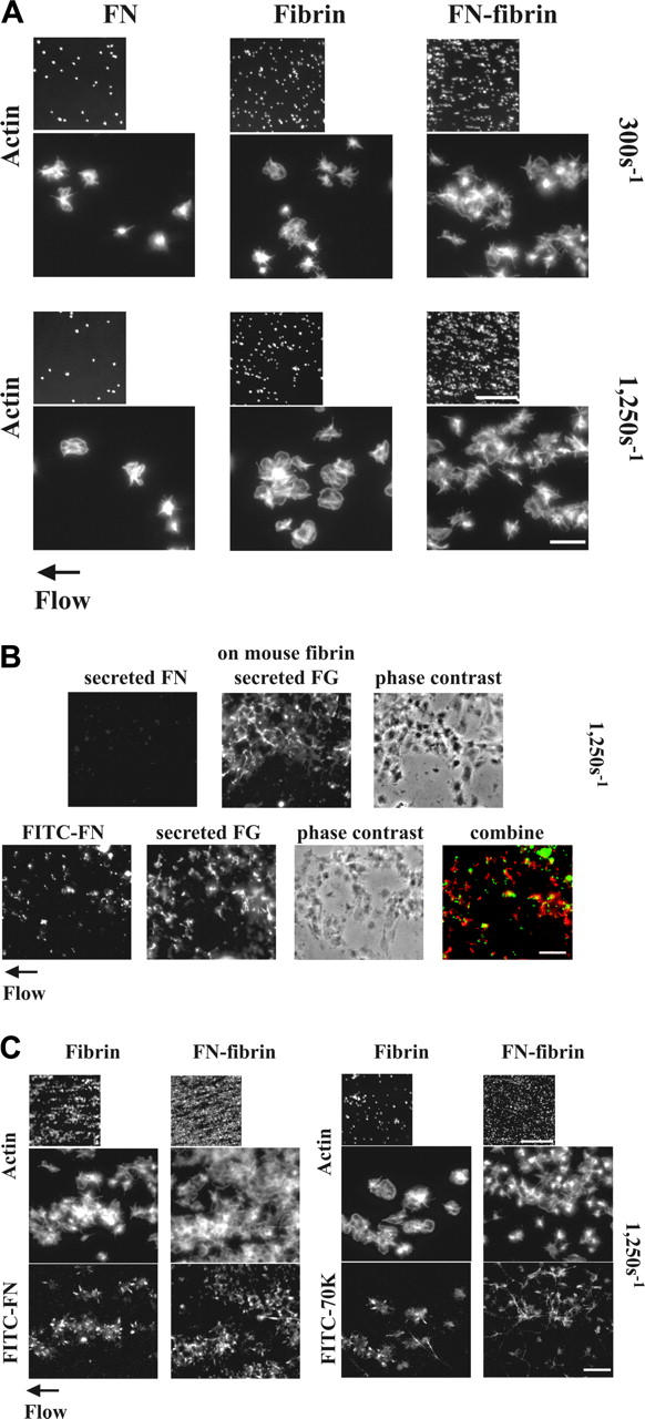
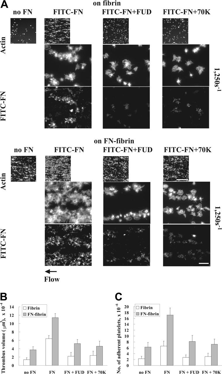
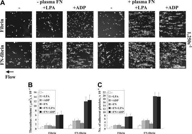
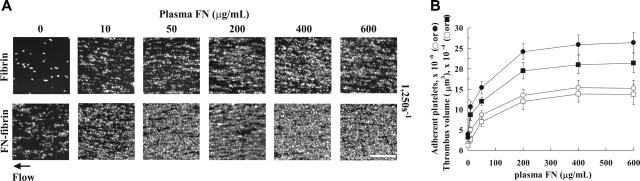
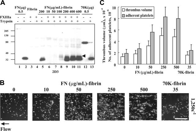
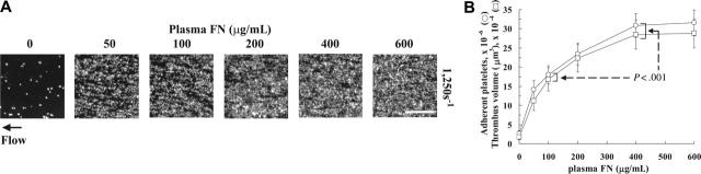
To learn how plasma fibronectin stabilizes platelet-rich thrombi in injured mesenteric arterioles of mice, we studied the impact of plasma fibronectin on platelet thrombus formation ex vivo in a parallel flow chamber. Thrombi were greater on surfaces coated with fibrin cross-linked to fibronectin by activated factor XIII than on surfaces coated with fibrin lacking cross-linked fibronectin or with fibronectin alone. Platelet thrombi were even greater when plasma fibronectin was perfused with platelets, resulting in deposition of the perfused fibronectin in platelet thrombi. The effect of perfused fibronectin on thrombogenesis was lost if fibronectin deposition was blocked by coperfusion with the N-terminal 70-kDa fragment of fibronectin or a peptide based on the functional upstream domain of protein F1 of Streptococcus pyogenes. Increases in thrombus formation were dependent on a platelet activator such as lysophosphatidic acid, amount of fibronectin cross-linked to fibrin, and concentration of fibronectin in the perfusate. The dependency of fibronectin concentration extended into the range of fibronectin concentrations associated with increased risk of coronary artery disease. At such concentrations, the 2 mechanisms for insolubilization of plasma fibronectin-cross-linking to fibrin and assembly by adherent and aggregating platelets-synergize to result in many-fold enhancement of platelet thrombus formation.


Figures

Figure 1.
Figure 1.
Adhesion and aggregation of platelets on fibrin and fibronectin-fibrin incorporation of soluble fibronectin or 70K fragment into platelet thrombi and effect of perfused fibronectin on the formation of platelet thrombi under shear conditions. (A) A suspension of platelets and red blood cells was perfused through a flow chamber opposed to a coverslip coated with fibrin or fibronectin-fibrin (FN-fibrin) at shear rates of 300 or 1250 s–1 for 5 minutes. Coverslips were taken out of the chamber and washed. Platelets were fixed, permeabilized, stained with rhodamine-phalloidin, and observed by epifluorescence microscopy. The small and large pictures were taken with × 10 (bar = 100 μm) and × 100 objectives (bar = 10 μm), respectively. (B-C) A suspension of platelets and red blood cells without soluble proteins (B) or premixed with 100 nM FITC-fibronectin or FITC-70K fragment (B-C) was perfused through a flow chamber opposed to a coverslip coated with human fibrin or fibronectin-fibrin, or mouse fibrin at a shear rate of 1250 s–1 for 5 minutes. (B) Platelets adhered to mouse fibrin-coated surfaces in the absence (top panel) or presence of FITC-fibronectin (bottom panel). Adherent platelets were fixed and incubated with a rabbit polyclonal antibody against human fibronectin and a mouse mAb against human fibrinogen, followed by incubation with FITC-conjugated anti–rabbit IgG and rhodamine-conjugated anti–mouse IgG antibody (top panel) or incubated only with mouse antifibrinogen antibody and rhodamine-conjugated antimouse IgG (bottom panel). The images in the lower panel were obtained by confocal microscopy and are from a 1-μm slice taken 4 μm above the plane of the coverslip. Bar = 10 μm. (C) Coverslips were taken out of the chamber after perfusion with FITC-fibronectin or FITC-70K fragment, and platelets were stained with rhodamine-phalloidin. Microscopy was performed as described. The small and large pictures were taken with × 10 (bar = 100 μm) and × 100 × (bar = 10 μm), respectively. The images of phalloidin-stained platelets should be compared with the bottom sections of panel A.
Figure 2.
Figure 2.
Effect of inhibition of deposition of perfused fibronectin on platelet thrombus formation under shear conditions. A suspension of platelets and red blood cells was premixed with 100 nM FITC-fibronectin (A) or unlabeled fibronectin (B-C) in the presence or absence of 1 μM FUD or 70K fragment and perfused through a flow chamber opposed to a coverslip or culture dish coated with fibrin or fibronectin-fibrin for 5 minutes at a shear rate of 1250 s–1. (A) Coverslips were taken out of the chamber, and microscopy was performed as described in Figure 1. The small and large pictures were taken with × 10 (bar = 100 μm) and × 100 objectives (bar = 10 μm), respectively. (B-C) Thrombus volumes and platelet numbers were measured as described in Table 1. Values represent the mean ± SD (n = 3 experiments).
Figure 3.
Figure 3.
Effects of LPA or ADP on platelet thrombus formation under shear conditions. A suspension of platelets and red blood cells in the presence or absence of 100 nM plasma fibronectin (FN) or 5 μM LPA or ADP was perfused through a flow chamber opposed to a coverslip or culture dish coated with fibrin or fibronectin-fibrin for 5 minutes at a shear rate of 1250 s–1. (A) Coverslips were taken out of the chamber, and microscopy was performed as described in Figure 1. Bar = 100 μm. (B-C) Thrombus volumes and platelet numbers were measured as described in Table 1. Values represent the mean ± SD (n = 3 experiments).
Figure 4.
Figure 4.
Effect of concentrations of plasma fibronectin in the perfusate on platelet thrombus formation under shear conditions. (A) A suspension of platelets and red blood cells was premixed with plasma fibronectin (FN), 10 to 600 μg/mL (20-1200 nM), and perfused through a flow chamber opposed to a coverslip or culture dish coated with fibrin or fibronectin-fibrin for 5 minutes at a shear rate of 1250 s–1. Coverslips were taken out of the chamber, and microscopy was performed as described in Figure 1. Bar = 100 μm. (B) Thrombus volumes (□, on fibrin or ▪ on fibronectin-fibrin) and platelet numbers (○, on fibrin or •, on fibronectin-fibrin) were measured as described in Table 1. Values represent the mean ± SD (n = 3-4 experiments).
Figure 5.
Figure 5.
Effect of amounts of fibronectin cross-linked to fibrin on platelet thrombus formation under shear conditions. (A) Fibrin clots incubated with different concentrations of fibronectin (FN) in the presence or absence of FXIIIa were digested with trypsin. The solubilized clots were electrophoresed under reduced conditions and immunoblotted with the 2D3, a monoclonal antibody against an epitope within the N-terminal 27-kDa tryptic fragment of fibronectin. Lane 1, 0.5 μg fibronectin; lane 2, 0.5 μg trypsin-digested fibronectin; lane 3, cross-linked fibrin lacking fibronectin; lane 4, trypsin-digested cross-linked fibrin lacking fibronectin; lane 5, trypsin digest of fibrin formed in the presence of fibronectin, 200 μg/mL, and absence of FXIIIa; lanes 6-11, trypsin digests of fibrin formed in the presence of fibronectin, 10, 50, 100, 200, 400, or 600 μg/mL, and FXIIIa, 5 μg/mL; lane 12, 0.5 μg 70K fragment; and lane 13, 0.5 μg trypsin-digested 70K fragment. Arrowhead indicates a 37-kDa band immunoblotted only in trypsin digests of fibrin cross-linked to fibronectin. The 27- and 37-kDa bands were not detected by nonimmune mouse IgG (not shown). (B) Fibronectin, 0 to 500 μg/mL, or 70K fragment, 35 μg/mL, was added to the mixture of human fibrinogen, 500 μg/mL; FXIII, 5 μg/mL; 2 mM CaCl2. The clot was formed by addition of thrombin, 1 U/mL, and after overnight incubation at 4°C, the bulk fibrin clots was removed. A suspension of platelets and red blood cells was perfused through a flow chamber opposed to a coverslip or culture dish coated with fibrin or fibrin cross-linked to different amounts of fibronectin or 70K fragment for 5 minutes at a shear rate of 1250 s–1. Platelet thrombus was visualized with rhodamine-phalloidin as described in Table 1. Bar = 100 μm. (C) Thrombus volumes and platelet numbers were measured as described in Table 1. Numbers on x-axis represent the concentration (μg/mL) of fibronectin or 70K fragment present during formation of a fibrin matrix. Values represent the mean ± SD (n = 3 experiments).
Figure 6.
Figure 6.
Effect of matched increases in the concentration of plasma fibronectin present during formation of fibrin matrices and in the perfusate on platelet thrombus formation under shear conditions. (A) Fibronectin (FN), 0 to 600 μg/mL, was present during formation of fibronectin-fibrin clots. The same concentration of plasma fibronectin was included in the perfusate. After 5 minutes, perfusion at a wall shear rate of 1250 s–1 coverslips were taken out of the chamber and washed. Microscopy was performed as described in Figure 1. Bar = 100 μm. (B) Thrombus volumes (□) and platelet numbers (○) were measured as described in Table 1. Values represent the mean ± SD (n = 4 experiments). The values at fibronectin concentrations of 400 and 600 μg/mL were significantly different from values at 100 μg/mL (P < .001 in the Dunnett test after ANOVA).

Similar articles

Cited by

References

    1. Mosher D. Fibronectin. San Diego, CA: Academic Press; 1989.
    1. Hynes R. Fibronectins. New York, NY: Springer-Verlag; 1990.
    1. Mosher DF. Fibronectin. Prog Hemost Thromb. 1980;5: 111-151. - PubMed
    1. Ginsberg MH, Plow EF. Fibronectin: a contender in platelet adhesive function. Academic Press, Harcourt Brace Jovanovich, Publishers; San Diego, CA: 1989.
    1. Sakai T, Johnson KJ, Murozono M, et al. Plasma fibronectin supports neuronal survival and reduces brain injury following transient focal cerebral ischemia but is not essential for skin-wound healing and hemostasis. Nat Med. 2001;7: 324-330. - PubMed

Publication types

MeSH terms