. 2006 May 1;107(9):3727-32.
doi: 10.1182/blood-2005-06-2259. Epub 2006 Jan 3.

TLR4-dependent hepcidin expression by myeloid cells in response to bacterial pathogens

Affiliations

TLR4-dependent hepcidin expression by myeloid cells in response to bacterial pathogens

Carole Peyssonnaux et al. Blood. .

Abstract

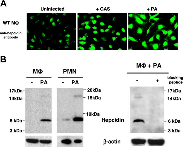
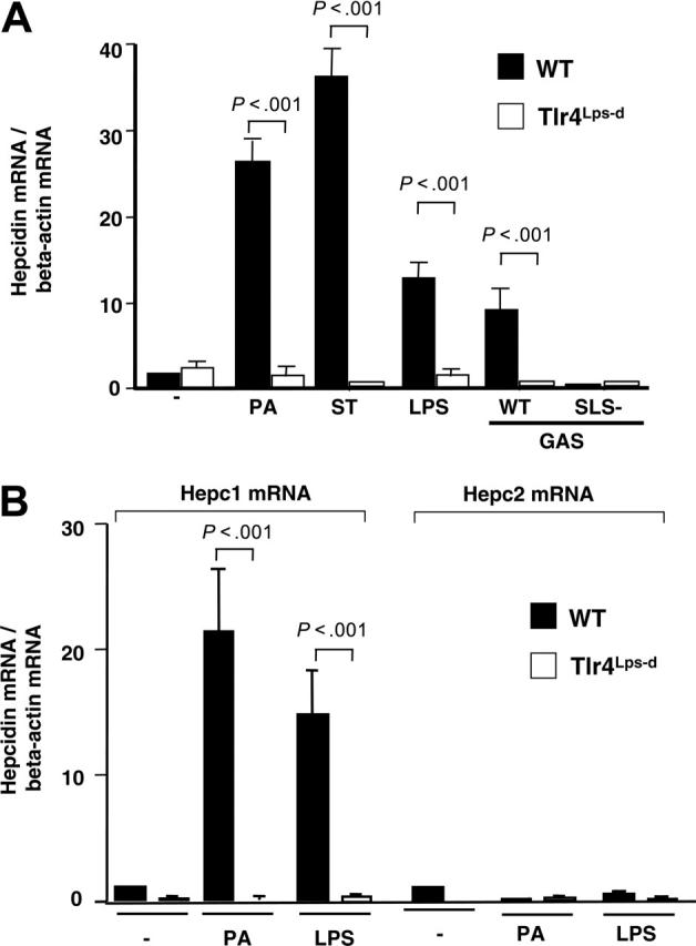
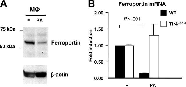
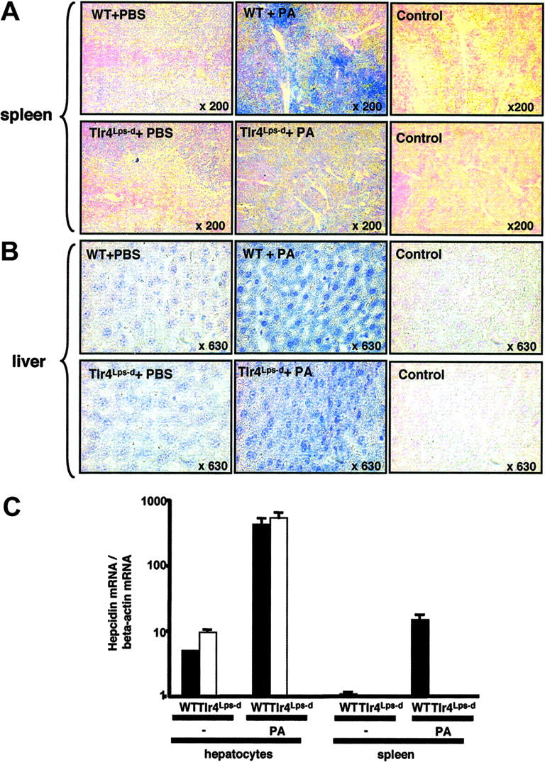
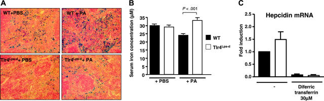
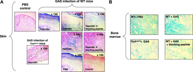
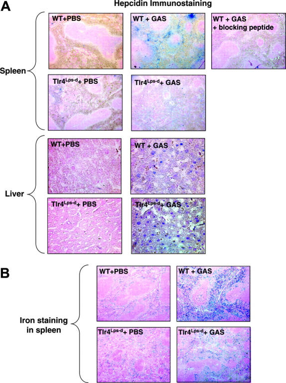
Hepcidin is an antimicrobial peptide secreted by the liver during inflammation that plays a central role in mammalian iron homeostasis. Here we demonstrate the endogenous expression of hepcidin by macrophages and neutrophils in vitro and in vivo. These myeloid cell types produced hepcidin in response to bacterial pathogens in a toll-like receptor 4 (TLR4)-dependent fashion. Conversely, bacterial stimulation of macrophages triggered a TLR4-dependent reduction in the iron exporter ferroportin. In vivo, intraperitoneal challenge with Pseudomonas aeruginosa induced TLR4-dependent hepcidin expression and iron deposition in splenic macrophages, findings mirrored in subcutaneous infection with group A Streptococcus where hepcidin induction was further observed in neutrophils migrating to the tissue site of infection. Hepcidin expression in cultured hepatocytes or in the livers of mice infected with bacteria was independent of TLR4, suggesting the TLR4-hepcidin pathway is restricted to myeloid cell types. Our findings identify endogenous myeloid cell hepcidin production as a previously unrecognized component of the host response to bacterial pathogens.


Figures

Figure 1.
Figure 1.
Hepcidin production is induced in neutrophils and macrophages by bacterial pathogens. (A) Immunofluorescence using anti-mouse hepcidin antibody on normal murine macrophages at baseline and upon exposure to Group A Streptococcus (GAS) or P aeruginosa (PA); magnification × 630. (B) Western blot analysis of hepcidin in murine macrophages and neutrophils at baseline and upon PA exposure. Addition of 10 μg blocking hepcidin peptide per milliliter of antibody was used to check for the specificity of the anti-mouse hepcidin antibody.
Figure 2.
Figure 2.
TLR4 dependency of bacterial-induced hepcidin mRNA expression by neutrophils and macrophages. (A) Real-time PCR for hepcidin mRNA on WT (CH3/HeouJ) and Tlr4Lps-d (CH3/HeJ) murine macrophages stimulated with P aeruginosa (PA), S typhimurium (ST), LPS, or Group A Streptococcus (GAS) producing or lacking the cytotoxin streptolysin S (SLS). Hepcidin mRNA was normalized to the expression level of β-actin. Quantitative assays performed in triplicate and representative of 3 repeated experiments. (B) Real-time PCR for hepcidin-1 and hepcidin-2 mRNA in WT (CH3/HeouJ) and Tlr4Lps-d (CH3/HeJ) macrophages stimulated with PA or LPS. Data are shown as mean of values ± standard deviation (SD).
Figure 3.
Figure 3.
TLR4-dependent ferroportin suppression by neutrophils and macrophages exposed to bacterial pathogens. (A) Western blot of ferroportin production in murine bone marrow derived macrophages stimulated with PA. (B) Real-time PCR for ferroportin mRNA using WT and Tlr4Lps-d bone marrow derived macrophages infected with PA. Quantitative assays performed in triplicate and representative of 3 repeated experiments. Data are shown as mean of values ± SD.
Figure 4.
Figure 4.
Hepcidin expression in spleen and liver of mice upon systemic infection. Immunohistochemistry using anti-mouse hepcidin antibodies on spleens (A) and livers (B) of mice 4 hours after intraperitoneal challenge with Pseudomonas aeruginosa or PBS control. (C) Real-time PCR for hepcidin-1 mRNA in liver and spleen of WT (CH3/HeouJ) and Tlr4Lps-d (CH3/HeJ) mice challenged for 4 hours with PA. Data are shown as mean ± SD
Figure 5.
Figure 5.
Splenic iron and measure of hypoferremia in WT and Tlr4Lps-d mice upon systemic infection. (A) Iron staining of splenic sections by Perl blue method 24 hour after intraperitoneal challenge with P aeruginosa (PA) or PBS control. (B) Serum iron measurement in WT and Tlr4Lps-d Tlr4Lps-d challenged with PA. (C) Hepcidin mRNA levels in WT and Tlr4Lps-d macrophages upon exposure to iron in the form of diferric transferrin.
Figure 6.
Figure 6.
Myeloid cell hepcidin expression in a subcutaneous model of infection. (A) Immunohistochemistry using anti-mouse hepcidin and anti-polymorphonuclear leukocyte (PMN) antibodies on skin of WT and Tlr4Lps-d mice challenged with group A Streptococcus (GAS); controls include omission of primary antibody and addition of blocking peptide to the hepcidin antibody. (B) Immunohistochemistry using anti-mouse hepcidin antibodies on bone marrow of mice challenged with GAS; control corresponds to the addition of hepcidin blocking peptide to the hepcidin antibody. Magnification, × 400.
Figure 7.
Figure 7.
Hepcidin expression in liver and spleen of mice infected with GAS and iron staining. (A) Immunohistochemistry using anti-mouse hepcidin antibodies on livers and spleens of mice challenged with group A Streptococcus (GAS), magnification × 50 (spleen) and × 400 (liver). (B) Iron staining of splenic sections by Perl blue method following similar mouse challenge.

References

    1. Ganz T. Hepcidin—a regulator of intestinal iron absorption and iron recycling by macrophages. Best Pract Res Clin Haematol. 2005;18: 171-182. - PubMed
    1. Krause A, Neitz S, Magert HJ, et al. LEAP-1, a novel highly disulfide-bonded human peptide, exhibits antimicrobial activity. FEBS Lett. 2000; 480: 147-150. - PubMed
    1. Park CH, Valore EV, Waring AJ, Ganz T. Hepcidin, a urinary antimicrobial peptide synthesized in the liver. J Biol Chem. 2001;276: 7806-7810. - PubMed
    1. Nicolas G, Bennoun M, Devaux I, et al. Lack of hepcidin gene expression and severe tissue iron overload in upstream stimulatory factor 2 (USF2) knockout mice. Proc Natl Acad Sci U S A. 2001; 98: 8780-8785. - PMC - PubMed
    1. Nicolas G, Bennoun M, Porteu A, et al. Severe iron deficiency anemia in transgenic mice expressing liver hepcidin. Proc Natl Acad Sci U S A. 2002;99: 4596-4601. - PMC - PubMed

Publication types

MeSH terms