. 2006 Jan;72(1):71-7.
doi: 10.1128/AEM.72.1.71-77.2006.

Improvement of alpha-amylase production by modulation of ribosomal component protein S12 in Bacillus subtilis 168

Affiliations

Improvement of alpha-amylase production by modulation of ribosomal component protein S12 in Bacillus subtilis 168

Kazuhiko Kurosawa et al. Appl Environ Microbiol. 2006 Jan.

Abstract

The capacity of ribosomal modification to improve antibiotic production by Streptomyces spp. has already been demonstrated. Here we show that introduction of mutations that produce streptomycin resistance (str) also enhances alpha-amylase (and protease) production by a strain of Bacillus subtilis as estimated by measuring the enzyme activity. The str mutations are point mutations within rpsL, the gene encoding the ribosomal protein S12. In vivo as well as in vitro poly(U)-directed cell-free translation systems showed that among the various rpsL mutations K56R (which corresponds to position 42 in E. coli) was particularly effective at enhancing alpha-amylase production. Cells harboring the K56R mutant ribosome exhibited enhanced translational activity during the stationary phase of cell growth. In addition, the K56R mutant ribosome exhibited increased 70S complex stability in the presence of low Mg2+ concentrations. We therefore conclude that the observed increase in protein synthesis activity by the K56R mutant ribosome reflects increased stability of the 70S complex and is responsible for the increase in alpha-amylase production seen in the affected strain.


Figures

FIG. 1.
FIG. 1.
Alignment of amino acid sequences of the ribosomal S12 proteins from several bacteria and location of mutations that confer resistance to streptomycin. Data are from the present work and references , and . Bs, Sc, and Ec represent Bacillus subtilis, Streptomyces coelicolor, and Escherichia coli, respectively. Asterisks indicate identical amino acids.
FIG. 2.
FIG. 2.
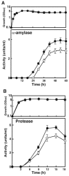
(A) Time course of growth (upper panel) and α-amylase production (lower panel) by B. subtilis in NG medium at 45°C. (B) Time course of growth and protease production in PM medium at 45°C. The values shown in each panel are the means of the results from three independent experiments. Error bars represent standard deviations. Symbols: ○, 168 (parental strain); •, Smr mutant WL1 (rpsL1). OD660, optical density at 660 nm.
FIG. 3.
FIG. 3.
Comparison of α-amylase production in the parental strain (168) and rpsL mutant strains (WL1 to WL15). Cultivation was carried out in NG medium for 54 h at 45°C. The values are means of the results from four independent experiments. Error bars represent standard deviations.
FIG. 4.
FIG. 4.
Capacity of wild-type (168) and mutant (WL1 and WL2) strains to synthesize proteins during the indicated growth phases. Incorporation of [3H]leucine into the total protein fraction was determined as described in Materials and Methods. The values are the means of the results from three independent experiments. Error bars represent standard deviations. OD600, optical density at 600 nm.
FIG. 5.
FIG. 5.
In vitro translation activities of ribosomes harvested from cells during the indicated growth phases. Strains were grown in NG medium at 45°C. Samples were taken during the indicated growth phases, and in vitro translation was carried out using a poly(U)-directed cell-free translation system (see Materials and Methods). The values are the means of the results from three independent experiments. Error bars represent standard deviations.
FIG. 6.
FIG. 6.
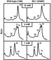
Sucrose gradient analysis of ribosome fractions. The patterns of dissociation of the 70S complex into 30S and 50S subunits at different concentrations of Mg2+ were determined using washed ribosomes harvested from cells grown to mid-exponential phase (2 h) in NG medium (see Materials and Methods). Asterisks indicate the peaks for the intermediates.

Similar articles

Cited by

References

    1. Ayusawa, D., Y. Yoneda, K. Yamane, and B. Maruo. 1975. Pleiotropic phenomena in autolytic enzyme(s) content, flagellation, and simultaneous hyperproduction of extracellular α-amylase and protease in a Bacillus subtilis mutant. J. Bacteriol. 124:459-469. - PMC - PubMed
    1. Bertoldo, C., and G. Antranikian. 2002. Starch-hydrolyzing enzymes from thermophilic archaea and bacteria. Curr. Opin. Chem. Biol. 6:151-160. - PubMed
    1. Carter, A. P., W. M. Clemons, D. E. Brodersen, R. J. Morgan-Warren, B. T. Wimberly, and V. Ramakrishnan. 2000. Functional insights from the structure of the 30S ribosomal subunit and its interactions with antibiotics. Nature 407:340-348. - PubMed
    1. Cundliffe, E. 1990. Recognition sites for antibiotics within rRNA, p. 479-490. In A. D. W. E. Hill, R. A. Garrett, P. B. Moore, D. Schlessinger, and J. R. Warner (ed.), The ribosome: structure, function, and evolution. American Society for Microbiology, Washington, D. C.
    1. Fujimoto, Z., K. Takase, N. Doui, M. Momma, T. Matsumoto, and H. Mizuno. 1998. Crystal structure of a catalytic-site mutant α-amylase from Bacillus subtilis complexed with maltopentaose. J. Mol. Biol. 277:393-407. - PubMed

Publication types

MeSH terms

LinkOut - more resources