

Efficient and stable display of functional proteins on bacterial magnetic particles using mms13 as a novel anchor molecule
- PMID: 16391079
- PMCID: PMC1352229
- DOI: 10.1128/AEM.72.1.465-471.2006
Efficient and stable display of functional proteins on bacterial magnetic particles using mms13 as a novel anchor molecule
Abstract
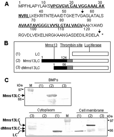
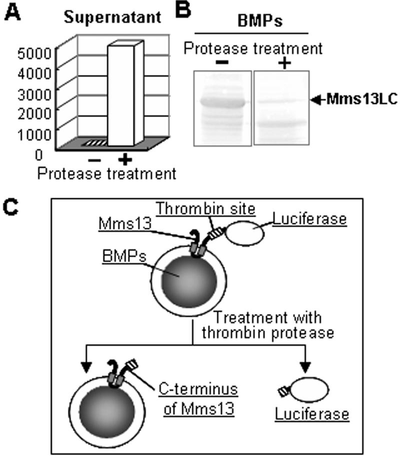
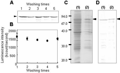
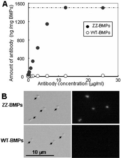
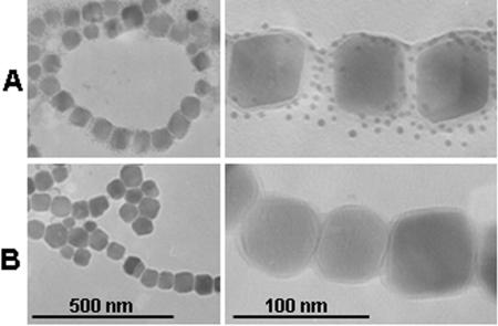
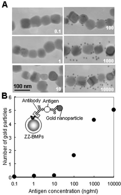
Magnetic particles are increasingly used for various biomedical applications because they are easy to handle and separate from biological samples. In this work, a novel anchor molecule was used for targeted protein display onto magnetic nanoparticles. The magnetic bacterium Magnetospirillum magneticum AMB-1 synthesizes intracellular bacterial magnetic particles (BMPs) covered with a lipid bilayer membrane. In our recent research, an integral BMP membrane protein, Mms13, was isolated and used as an anchor molecule to display functional proteins onto BMPs. The anchoring properties of Mms13 were confirmed by luciferase fusion studies. The C terminus of Mms13 was shown to be expressed on the surface of BMPs, and Mms13 was bound to magnetite directly and tightly permitting stable localization of a large protein, luciferase (61 kDa), on BMPs. Consequently, luminescence intensity obtained from BMPs using Mms13 as an anchor molecule was >400 or 1,000 times higher than Mms16 or MagA, which previously were used as anchor molecules. Furthermore, the immunoglobulin G-binding domain of protein A (ZZ) was displayed uniformly on BMPs using Mms13, and antigen was detected by transmission electron microscopy using antibody-labeled gold nanoparticles on a single BMP displaying the ZZ-antibody complex. The results of this study demonstrated the utility of Mms13 as a molecular anchor, which will facilitate the assembly of other functional proteins onto BMPs in the near feature.
Figures
Similar articles
-
Development of efficient expression system for protein display on bacterial magnetic particles.Biochem Biophys Res Commun. 2005 Dec 30;338(4):1678-81. doi: 10.1016/j.bbrc.2005.10.148. Epub 2005 Nov 2. Biochem Biophys Res Commun. 2005. PMID: 16288989
-
Design, expression and characterization of single chain Fv, Mms13 and the single chain Fv‑mms13 fusion protein.Mol Med Rep. 2015 Jul;12(1):1213-8. doi: 10.3892/mmr.2015.3561. Epub 2015 Mar 27. Mol Med Rep. 2015. PMID: 25824464
-
Preparation of luciferase-bacterial magnetic particle complex by artificial integration of MagA-luciferase fusion protein into the bacterial magnetic particle membrane.Biotechnol Bioeng. 2002 Mar 20;77(6):614-8. doi: 10.1002/bit.10114. Biotechnol Bioeng. 2002. PMID: 11807756
-
Bioengineering of bacterial magnetic particles and their applications in biotechnology.Recent Pat Biotechnol. 2010 Nov;4(3):214-25. doi: 10.2174/187220810793611455. Recent Pat Biotechnol. 2010. PMID: 21171958 Review.
-
Molecular analysis of magnetotactic bacteria and development of functional bacterial magnetic particles for nano-biotechnology.Trends Biotechnol. 2007 Apr;25(4):182-8. doi: 10.1016/j.tibtech.2007.02.002. Epub 2007 Feb 15. Trends Biotechnol. 2007. PMID: 17306901 Review.
Cited by
-
In vivo display of a multisubunit enzyme complex on biogenic magnetic nanoparticles.Appl Environ Microbiol. 2009 Dec;75(24):7734-8. doi: 10.1128/AEM.01640-09. Epub 2009 Oct 16. Appl Environ Microbiol. 2009. PMID: 19837839 Free PMC article.
-
Magnetosome expression of functional camelid antibody fragments (nanobodies) in Magnetospirillum gryphiswaldense.Appl Environ Microbiol. 2011 Sep;77(17):6165-71. doi: 10.1128/AEM.05282-11. Epub 2011 Jul 15. Appl Environ Microbiol. 2011. PMID: 21764974 Free PMC article.
-
From invagination to navigation: The story of magnetosome-associated proteins in magnetotactic bacteria.Protein Sci. 2016 Feb;25(2):338-51. doi: 10.1002/pro.2827. Epub 2015 Nov 3. Protein Sci. 2016. PMID: 26457474 Free PMC article. Review.
-
The major magnetosome proteins MamGFDC are not essential for magnetite biomineralization in Magnetospirillum gryphiswaldense but regulate the size of magnetosome crystals.J Bacteriol. 2008 Jan;190(1):377-86. doi: 10.1128/JB.01371-07. Epub 2007 Oct 26. J Bacteriol. 2008. PMID: 17965152 Free PMC article.
-
Relevant uses of surface proteins--display on self-organized biological structures.Microb Biotechnol. 2012 Mar;5(2):188-202. doi: 10.1111/j.1751-7915.2011.00293.x. Epub 2011 Sep 9. Microb Biotechnol. 2012. PMID: 21906264 Free PMC article. Review.
References
-
- Arakaki, A., J. Webb, and T. Matsunaga. 2003. A novel protein tightly bound to bacterial magnetic particles in Magnetospirillum magneticum strain AMB-1. J. Biol. Chem. 278:8745-8750. - PubMed
-
- Clackson, T., H. R. Hoogenboom, A. D. Griffiths, and G. Winter. 1991. Making antibody fragments using phage display libraries. Nature 352:624-628. - PubMed
-
- Gao, X., Y. Cui, R. M. Levenson, L. W. Chung, and S. Nie. 2004. In vivo cancer targeting and imaging with semiconductor quantum dots. Nat. Biotechnol. 22:969-976. - PubMed
-
- Goldman, E. R., G. P. Anderson, P. T. Tran, H. Mattoussi, P. T. Charles, and J. M. Mauro. 2002. Conjugation of luminescent quantum dots with antibodies using an engineered adaptor protein to provide new reagents for fluoroimmunoassays. Anal. Chem. 74:841-847. - PubMed
Publication types
MeSH terms
Substances
LinkOut - more resources
Full Text Sources
Other Literature Sources
