

Replication and long-term persistence of bovine and human strains of Mycobacterium avium subsp. paratuberculosis within Acanthamoeba polyphaga
- PMID: 16391127
- PMCID: PMC1352277
- DOI: 10.1128/AEM.72.1.854-859.2006
Replication and long-term persistence of bovine and human strains of Mycobacterium avium subsp. paratuberculosis within Acanthamoeba polyphaga
Abstract
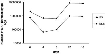
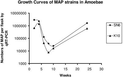
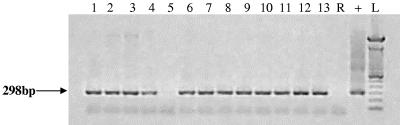
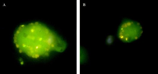
Free-living protists are ubiquitous in the environment and form a potential reservoir for the persistence of animal and human pathogens. Mycobacterium avium subsp. paratuberculosis is the cause of Johne's disease, a systemic infection accompanied by chronic inflammation of the intestine that affects many animals, including primates. Most humans with Crohn's disease are infected with this chronic enteric pathogen. Subclinical infection with M. avium subsp. paratuberculosis is widespread in domestic livestock. Infected animals excrete large numbers of robust organisms into the environment, but little is known about their ability to replicate and persist in protists. In the present study we fed laboratory cultures of Acanthamoeba polyphaga with bovine and human strains of M. avium subsp. paratuberculosis. Real-time PCR showed that the numbers of the pathogens fell over the first 4 to 8 days and recovered by 12 to 16 days. Encystment of the amoebic cultures after 4 weeks resulted in a 2-log reduction in the level of M. avium subsp. paratuberculosis, which returned to the original level by 24 weeks. Extracts of resection samples of human gut from 39 patients undergoing abdominal surgery were fed to cultures of A. polyphaga. M. avium subsp. paratuberculosis detected by nested IS900 PCR with amplicon sequencing and visualized by IS900 in situ hybridization and auramine-rhodamine staining was found in cultures derived from 13 of the patients and was still present in the cultures after almost 4 years of incubation. Control cultures were negative. M. avium subsp. paratuberculosis has the potential for long-term persistence in environmental protists.
Figures
Similar articles
-
Evaluation of in situ methods used to detect Mycobacterium avium subsp. paratuberculosis in samples from patients with Crohn's disease.J Clin Microbiol. 2006 Aug;44(8):2942-50. doi: 10.1128/JCM.00585-06. J Clin Microbiol. 2006. PMID: 16891515 Free PMC article.
-
Detection and verification of Mycobacterium avium subsp. paratuberculosis in fresh ileocolonic mucosal biopsy specimens from individuals with and without Crohn's disease.J Clin Microbiol. 2003 Jul;41(7):2915-23. doi: 10.1128/JCM.41.7.2915-2923.2003. J Clin Microbiol. 2003. PMID: 12843021 Free PMC article.
-
Mycobacterium avium subspecies paratuberculosis infection in cases of irritable bowel syndrome and comparison with Crohn's disease and Johne's disease: common neural and immune pathogenicities.J Clin Microbiol. 2007 Dec;45(12):3883-90. doi: 10.1128/JCM.01371-07. Epub 2007 Oct 3. J Clin Microbiol. 2007. PMID: 17913930 Free PMC article.
-
Current perspectives on Mycobacterium avium subsp. paratuberculosis, Johne's disease, and Crohn's disease: a review.Crit Rev Microbiol. 2011 May;37(2):141-56. doi: 10.3109/1040841X.2010.532480. Epub 2011 Jan 22. Crit Rev Microbiol. 2011. PMID: 21254832 Review.
-
[Mycobacterium avium subsp. paratuberculosis in food and its relationship with Crohn's disease].Rev Argent Microbiol. 2007 Jan-Mar;39(1):57-68. Rev Argent Microbiol. 2007. PMID: 17585661 Review. Spanish.
Cited by
-
Long-term survival and virulence of Mycobacterium leprae in amoebal cysts.PLoS Negl Trop Dis. 2014 Dec 18;8(12):e3405. doi: 10.1371/journal.pntd.0003405. eCollection 2014 Dec. PLoS Negl Trop Dis. 2014. PMID: 25521850 Free PMC article.
-
Extensive environmental survey of free-living amoebae and their elusive association with Mycobacterium bovis or Mycobacterium avium subsp. paratuberculosis.FEMS Microbiol Ecol. 2025 Jan 7;101(1):fiae164. doi: 10.1093/femsec/fiae164. FEMS Microbiol Ecol. 2025. PMID: 39689919 Free PMC article.
-
"Candidatus Fokinia solitaria", a Novel "Stand-Alone" Symbiotic Lineage of Midichloriaceae (Rickettsiales).PLoS One. 2016 Jan 5;11(1):e0145743. doi: 10.1371/journal.pone.0145743. eCollection 2016. PLoS One. 2016. PMID: 26731731 Free PMC article.
-
Assessing the inactivation of Mycobacterium avium subsp. paratuberculosis during composting of livestock carcasses.Appl Environ Microbiol. 2013 May;79(10):3215-24. doi: 10.1128/AEM.03768-12. Epub 2013 Mar 15. Appl Environ Microbiol. 2013. PMID: 23503307 Free PMC article.
-
Evaluation of in situ methods used to detect Mycobacterium avium subsp. paratuberculosis in samples from patients with Crohn's disease.J Clin Microbiol. 2006 Aug;44(8):2942-50. doi: 10.1128/JCM.00585-06. J Clin Microbiol. 2006. PMID: 16891515 Free PMC article.
References
-
- Anderson, O. R. 2000. Abundance of terrestrial gymnamoebae at a northeastern U. S. site: a four-year study, including the El Nino winter of 1997-1998. J. Eukaryot. Microbiol. 47:148-155. - PubMed
-
- Barker, J., and M. R. Brown. 1994. Trojan horses of the microbial world: protozoa and the survival of bacterial pathogens in the environment. Microbiology 140:1253-1259. - PubMed
Publication types
MeSH terms
Substances
LinkOut - more resources
Full Text Sources
