. 2006 Jan 1;20(1):47-64.
doi: 10.1101/gad.1384406.

A pathway in quiescent cells that controls p27Kip1 stability, subcellular localization, and tumor suppression

Affiliations

A pathway in quiescent cells that controls p27Kip1 stability, subcellular localization, and tumor suppression

Arnaud Besson et al. Genes Dev. .

Abstract

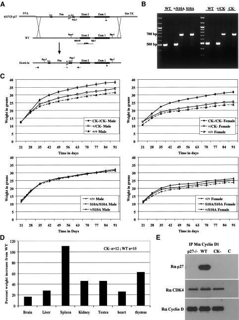
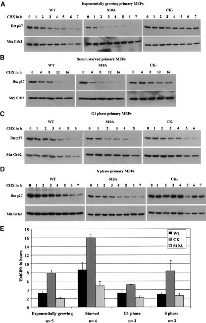
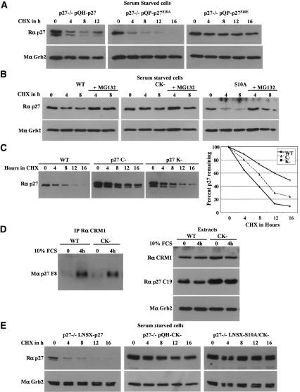
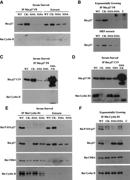
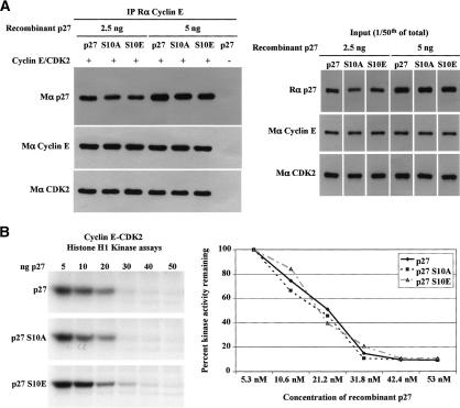
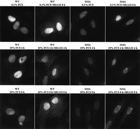
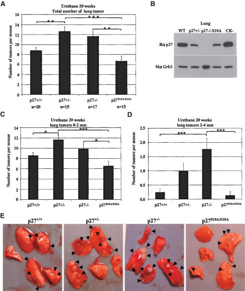
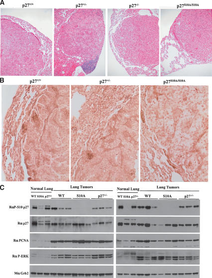
We have created two knock-in mouse models to study the mechanisms that regulate p27 in normal cells and cause misregulation of p27 in tumors: p27(S10A), in which Ser10 is mutated to Ala; and p27(CK-), in which point mutations abrogate the ability of p27 to bind cyclins and CDKs. These two mutant alleles identify steps in a pathway that controls the proteasomal degradation of p27 uniquely in quiescent cells: Dephosphorylation of p27 on Ser10 inhibits p27 nuclear export and promotes its assembly into cyclin-CDK complexes, which is, in turn, necessary for p27 turnover. We further show that Ras-dependent lung tumorigenesis is associated with increased phosphorylation on Ser10 and cytoplasmic mislocalization of p27. Indeed, we find that p27(S10A) is refractory to Ras-induced cytoplasmic translocation and that p27(S10A) mice are tumor resistant. Thus, phosphorylation of p27 on Ser10 is an important event in the regulation of the tumor suppressor function of p27.


Figures

Figure 1.
Figure 1.
Construction of p27 mutant mice. (A) Targeting strategy for generating p27 knock-in mice. Schematic of the targeting construct used to replace the wild-type p27 genomic region with the p27S10A or p27CK– alleles. The Southern probe as well as the PCR primers used for screening recombinant ES cells and genotyping transgenic mice are indicated. (B) Genotyping PCR. Using the primers shown in A, the wild-type allele gives a PCR product of ∼500 bp and the recombinant allele (either p27S10A or p27CK–) a 700-bp product. (C) Size increase of the p27CK–/CK– mice. Thirty animals per genotype were weighed weekly from weaning to 3 mo of age. Both male and female p27CK–/CK– mice are larger than their wild-type counterparts. The size of p27S10A/S10A mice was not significantly affected, although females were slightly larger. (D) Percent increases in the mean weight of individual organs from 12 p27CK–/CK– males compared with that of 10 wild-type mice. (E) p27CK– does not interact with cyclin D1. Cyclin D1 was immunoprecipitated from the indicated primary MEF extracts. Immunoprecipitates were resolved on SDS-PAGE and the membrane was probed for p27 (C19), stripped, and reprobed successively for CDK4 and cyclin D. The control lane (C) refers to an IP of wild-type extract with an irrelevant antibody.
Figure 2.
Figure 2.
Opposite effects of the p27CK– and p27S10A mutations on p27 levels and on p27 degradation following growth stimulation. (A) Tissue expression of p27 in p27+/+ (WT), p27S10A (S10A), p27–/–, and p27CK– (CK) mice. Equal amounts of tissue extracts (spleen, brain, uterus, liver) were probed successively with a polyclonal p27 antibody (C19) and with a monoclonal anti-Grb2 antibody to evaluate protein loading. (B) Lack of Ser10 phosphorylation of the p27S10A mutant. Brain extracts were probed successively with a phospho-Ser10 p27-specific antibody, and for p27 and Grb2 (loading control). (C) p27S10A and p27CK– levels in primary MEFs in different culture conditions. MEFs were either exponentially growing (Exp growing), serum starved for 72 h (0.1% FCS), or kept confluent for 24 h (Confluent). The membrane was probed as in B. (D) p27S10A and p27CK– have oppositely altered kinetics of degradation following serum stimulation. Wild-type, p27S10A, and p27CK– primary MEFs were serum starved for 72 h and replated in the presence of 10% serum. Membranes were probed as in A. (E) Flow cytometric analyses of the cell cycle of wild-type, p27S10A, and p27CK– primary MEFs. Parallel cell cultures as in D were treated similarly and collected for flow cytometry.
Figure 3.
Figure 3.
p27S10A is unstable in quiescent cells, while p27CK– is stabilized in G0 and S phases. p27 half-life determination in exponentially growing (A), serum-starved for 72 h (B), G1-phase (C), and S-phase (D) wild-type (WT), p27S10A (S10A), and p27CK– (CK) primary MEFs. (AD) Primary MEFs in the indicated culture conditions were treated with 25 μg/mL cycloheximide (CHX) and collected at the indicated time. A polyclonal p27 antibody (C19), and a monoclonal Grb2 (loading control) were used for immunoblotting. (E) Summary of p27 half-life data from several experiments: exponentially growing (n = 5), serum starved (n = 4), G1 phase (n = 3), and S phase (n = 3). For each experiment, p27 immunoblots were quantified and the amount of p27 expressed as percent of p27 remaining from time zero. Half-lives were determined from the individual graphs by drawing a “best fit” curve for each cell type and condition. (*) Note that the p27CK– half-life in S phase is underestimated in the graph as essentially no turnover occurred during the time frame in one experiment (i.e., T½ ≫ 7 h), and this experiment therefore could not be included in the calculation of half-life.
Figure 4.
Figure 4.
The instability of p27S10A depends on its ability to interact with cyclin–CDK complexes. (A) A Ser10-phosphomimetic mutant (p27S10E) is stable in quiescent cells. p27-null immortalized MEFs were retrovirally infected with either wild-type p27 (p27–/–pQH-p27), p27S10A (p27–/–pQP-p27S10A), or p27S10E (p27–/–pQP-p27S10E). After selection of infected MEFs, cells were serum starved, treated with CHX, and processed as described in Figure 3. (B) p27 degradation is proteasome-dependent in quiescent cells. Wild type (WT), p27S10A (S10A), and p27CK– (CK) primary MEFs were serum starved for 72 h and treated with 25 μg/mL CHX or with both CHX and 10 μM MG132 for the indicated time. Samples were processed as in Figure 3. (C) Cyclin interaction is critical for p27 turnover in G0. The half-lives of wild-type p27, p27C– (p27–/–pQP-p27C–), and p27K– (p27–/–pQP-p27K–) were determined as in A. (D) p27CK– can still interact with CRM1. CRM1 was immunoprecipitated from extracts prepared from primary wild-type or p27CK– MEFs either serum starved for 72 h, or starved and stimulated for 4 h with 10% serum. Immunoprecipitates were resolved on SDS-PAGE and probed for p27. A fraction of the extracts was used for Western blotting to determine the amounts of CRM1, p27, and Grb2 (loading control) present in these cells. (E) Stabilization of a p27S10A mutant that cannot bind cyclins and CDKs (p27S10A/CK–) in quiescent cells. p27-null MEFs were retrovirally infected with either wild-type p27 (p27–/–LNSX-p27), p27CK– (p27–/–pQH-CK), or p27S10A/CK– (p27–/–LNSX-S10A/CK), and treated as in A.
Figure 5.
Figure 5.
Most p27S10A is bound to cyclin–CDK complexes. IPs in primary MEF protein extracts. (A,B) IP with a monoclonal p27 antibody (p27 F8) that immunoprecipitates only free p27 in serum-starved (A) or exponentially growing (B) cells. The amounts of p27 present in the cell extracts used for IP are also indicated. Immunoprecipitates were resolved on SDS-PAGE, and membranes were probed with a polyclonal p27 antibody (C19). In A, the membrane was stripped and reprobed with a polyclonal antibody to cyclin D. (C) Same as A, except that an additional extract from p27–/– cells expressing a p27S10A/CK– mutant was used for IP. (D) Same as C with IPs performed with a polyclonal p27 antibody (C19) that immunoprecipitates all forms of p27. (E,F) IPs with a monoclonal cyclin D1 antibody in serum-starved (E) or exponentially growing (F) primary MEFs. Membranes were probed successively with polyclonal antibodies to phospho-Ser10 p27, p27, CDK4, and cyclin D. For F, the cell extracts used were the same as in B. In B and F, the control lane (C) corresponds to an IP of wild-type extract performed with an irrelevant antibody.
Figure 6.
Figure 6.
p27S10A, p27S10E, and wild-type p27 bind to and inhibit cyclin E–CDK2 complexes in vitro equivalently. (A) Recombinant wild-type p27, p27S10A, and p27S10E bind cyclin E–CDK2 complexes with similar affinities. The indicated amounts of recombinant p27 proteins were incubated with cyclin E–CDK2 complexes expressed in SF21 cells. Cyclin E was then immunoprecipitated, and the amount of p27 coprecipitated was determined by Western blotting; amounts of cyclin E and CDK2 immunoprecipitated are also shown. The right panel shows the starting amounts of proteins used in the binding assay. (B) Recombinant wild-type p27, p27S10A, and p27S10E have similar abilities to inhibit cyclin E–CDK2 complexes in in vitro histone H1 kinase assays. The graph indicates the percent of kinase activity remaining normalized to the control reaction, where there is no inhibition by p27.
Figure 7.
Figure 7.
Nuclear sequestration of p27S10A. Immunofluorescence with a monoclonal p27 antibody (clone 57; BD-Transduction Laboratories) in the presence or absence of 10 μM MG132 in wild-type and p27S10A MEFs either serum starved for 48 h (0.1% FCS), or starved and stimulated with 10% FCS for 4 h or 8 h. All images were taken with identical settings using a 100× lens, with the exposure time calculated on p27–/– MEFs processed identically so that the nonspecific staining of the antibody was not visible.
Figure 8.
Figure 8.
Decreased incidence and growth rate of urethane-induced lung tumors in p27S10A mice. (A) Total number of lung tumors in wild-type, p27+/–, p27–/–, and p27S10A mice 20 wk following a single dose of urethane. Lungs were dissected and macroscopic tumors were counted by visual inspection. The results were analyzed using a one-way analysis of variance with the Tukey-Kramer multiple comparison test. (***) p < 0.001; (**) p < 0.01; (*) p < 0.05. (B) p27S10A protein levels in the lung are similar to p27 levels in p27+/– mice. Lung extracts from wild-type (WT), p27+/–, p27–/–, p27S10A/S10A (S10A), and p27CK–/CK (CK) mice were resolved on SDS-PAGE, and the membrane was probed with a polyclonal p27 (C19) antibody, stripped, and reprobed with a monoclonal Grb2 antibody to evaluate protein loading. (C,D) Lung tumors were counted according to their size: small tumors (between 0 and 2 mm in diameter) (C), and large tumors (between 2 and 4 mm in diameter) (D). Statistical analyses were performed as in A. (E) Representative lungs for each mouse genotype following dissection. Tumors are indicated by arrows.
Figure 9.
Figure 9.
Nuclear localization of p27S10A is maintained in lung tumors. (A) Lung tumor histology. Five-micron-thick slices of paraffin-embedded lungs tissues from wild-type, p27+/–, p27–/–, and p27S10A/S10A mice were stained with eosin and hematoxylin. Tumors were classified as bronchioalveolar adenomas and papillary adenomas. (B) p27 immunohistochemistry of lung tumors and normal surrounding tissue. Five-micron-thick slices of paraffin-embedded lung tissues from wild-type, p27+/–, and p27S10A/S10A mice were stained using the mouse monoclonal p27 antibody (clone 57; BD-Transduction Laboratories). (C) Western blot analysis of normal lung tissue and individual tumors form wild-type, p27+/–, and p27S10A/S10A mice. Forty micrograms of proteins were loaded per well. Membranes were probed successively with polyclonal antibodies to phospho-Ser10 p27, p27, PCNA, phospho-ERK (activated), and with a monoclonal anti-Grb2 antibody for protein loading.
Figure 10.
Figure 10.
Unlike wild-type p27, p27S10A is resistant to activated Ras-induced cytoplasmic translocation. p27-null, wild-type, and p27S10A MEFs either expressing K-Ras V12 or not were serum starved for 72 h and stimulated with 10% serum in the presence or absence of MG132, fixed, and stained with a monoclonal antibody to p27. As in Figure 7, the exposure time was set on the p27–/– cells (left column) and kept constant to acquire all the other images, to take into account the nonspecific signal generated by the antibody.

Similar articles

Cited by

References

    1. Barbin A. 2000. Etheno-adduct-forming chemicals: From mutagenicity testing to tumor mutation spectra. Mutat. Res. 462: 55–69. - PubMed
    1. Besson A. and Yong, V.W. 2000. Involvement of p21(Waf1/Cip1) in protein kinase C α-induced cell cycle progression. Mol. Cell. Biol. 20: 4580–4590. - PMC - PubMed
    1. Besson A., Assoian, R.K., and Roberts, J.M. 2004a. Regulation of the cytoskeleton: An oncogenic function for CDK inhibitors? Nat. Rev. Cancer 4: 948–955. - PubMed
    1. Besson A., Gurian-West, M., Schmidt, A., Hall, A., and Roberts, J.M. 2004b. p27Kip1 modulates cell migration through the regulation of RhoA activation. Genes & Dev. 18: 862–876. - PMC - PubMed
    1. Boehm M., Yoshimoto, T., Crook, M.F., Nallamshetty, S., True, A., Nabel, G.J., and Nabel, E.G. 2002. A growth factor-dependent nuclear kinase phosphorylates p27Kip1 and regulates cell cycle progression. EMBO J. 21: 3390–3401. - PMC - PubMed

Publication types

MeSH terms