Cyclooxygenase-1-derived PGE2 promotes cell motility via the G-protein-coupled EP4 receptor during vertebrate gastrulation
- PMID: 16391234
- PMCID: PMC1356102
- DOI: 10.1101/gad.1374506
Cyclooxygenase-1-derived PGE2 promotes cell motility via the G-protein-coupled EP4 receptor during vertebrate gastrulation
Abstract
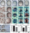
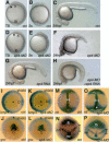
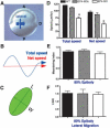
Gastrulation is a fundamental process during embryogenesis that shapes proper body architecture and establishes three germ layers through coordinated cellular actions of proliferation, fate specification, and movement. Although many molecular pathways involved in the specification of cell fate and polarity during vertebrate gastrulation have been identified, little is known of the signaling that imparts cell motility. Here we show that prostaglandin E(2) (PGE(2)) production by microsomal PGE(2) synthase (Ptges) is essential for gastrulation movements in zebrafish. Furthermore, PGE(2) signaling regulates morphogenetic movements of convergence and extension as well as epiboly through the G-protein-coupled PGE(2) receptor (EP4) via phosphatidylinositol 3-kinase (PI3K)/Akt. EP4 signaling is not required for proper cell shape or persistence of migration, but rather it promotes optimal cell migration speed during gastrulation. This work demonstrates a critical requirement of PGE(2) signaling in promoting cell motility through the COX-1-Ptges-EP4 pathway, a previously unrecognized role for this biologically active lipid in early animal development.
Figures


Similar articles
-
Prostaglandin Gbetagamma signaling stimulates gastrulation movements by limiting cell adhesion through Snai1a stabilization.Development. 2010 Apr;137(8):1327-37. doi: 10.1242/dev.045971. Development. 2010. PMID: 20332150 Free PMC article.
-
Prostaglandin E2 promotes lung cancer cell migration via EP4-betaArrestin1-c-Src signalsome.Mol Cancer Res. 2010 Apr;8(4):569-77. doi: 10.1158/1541-7786.MCR-09-0511. Epub 2010 Mar 30. Mol Cancer Res. 2010. PMID: 20353998 Free PMC article.
-
Prostaglandin E2-EP4 receptor promotes endothelial cell migration via ERK activation and angiogenesis in vivo.J Biol Chem. 2007 Jun 8;282(23):16959-68. doi: 10.1074/jbc.M701214200. Epub 2007 Mar 31. J Biol Chem. 2007. PMID: 17401137
-
Zebrafish gastrulation: cell movements, signals, and mechanisms.Int Rev Cytol. 2007;261:159-92. doi: 10.1016/S0074-7696(07)61004-3. Int Rev Cytol. 2007. PMID: 17560282 Review.
-
Gastrulation in zebrafish -- all just about adhesion?Curr Opin Genet Dev. 2006 Aug;16(4):433-41. doi: 10.1016/j.gde.2006.06.009. Epub 2006 Jun 23. Curr Opin Genet Dev. 2006. PMID: 16797963 Review.
Cited by
-
Discovering chemical modifiers of oncogene-regulated hematopoietic differentiation.Nat Chem Biol. 2009 Apr;5(4):236-43. doi: 10.1038/nchembio.147. Epub 2009 Jan 26. Nat Chem Biol. 2009. PMID: 19172146 Free PMC article.
-
Prostaglandin signalling regulates ciliogenesis by modulating intraflagellar transport.Nat Cell Biol. 2014 Sep;16(9):841-51. doi: 10.1038/ncb3029. Epub 2014 Aug 31. Nat Cell Biol. 2014. PMID: 25173977 Free PMC article.
-
Prostaglandin E2 regulates vertebrate haematopoietic stem cell homeostasis.Nature. 2007 Jun 21;447(7147):1007-11. doi: 10.1038/nature05883. Nature. 2007. PMID: 17581586 Free PMC article.
-
Zebrafish lipid metabolism: from mediating early patterning to the metabolism of dietary fat and cholesterol.Methods Cell Biol. 2011;101:111-41. doi: 10.1016/B978-0-12-387036-0.00005-0. Methods Cell Biol. 2011. PMID: 21550441 Free PMC article. Review.
-
NSAID-mediated cyclooxygenase inhibition disrupts ectodermal derivative formation in axolotl embryos.bioRxiv [Preprint]. 2025 Feb 15:2024.10.30.621122. doi: 10.1101/2024.10.30.621122. bioRxiv. 2025. Update in: Differentiation. 2025 May-Jun;143:100856. doi: 10.1016/j.diff.2025.100856. PMID: 39554061 Free PMC article. Updated. Preprint.
References
-
- Breyer R.M., Bagdassarian, C.K., Myers, S.A., and Breyer, M.D. 2001. Prostanoid receptors: Subtypes and signaling. Ann. Rev. Pharmacol. Toxicol. 41: 661–690. - PubMed
-
- Buchanan F.G., Wang, D., Bargiacchi, F., and DuBois, R.N. 2003. Prostaglandin E2 regulates cell migration via the intracellular activation of the epidermal growth factor receptor. J. Biol. Chem. 278: 35451–35457. - PubMed
-
- Carreira-Barbosa F., Concha, M.L., Takeuchi, M., Ueno, N., Wilson, S.W., and Tada, M. 2003. Prickle 1 regulates cell movements during gastrulation and neuronal migration in zebrafish. Development 130: 4037–4046. - PubMed
-
- Cha Y.I., Kim, S.H., Solnica-Krezel, L., and DuBois, R.N. 2005a. Cyclooxygenase-1 signaling is required for vascular tube formation during development. Dev. Biol. 282: 274–283. - PubMed
-
- Cha Y.I., Solnica-Krezel, L., and DuBois, R.N. 2005b. Fishing for prostanoids: Deciphering the developmental functions of cyclooxygenase-derived prostaglandins. Dev. Biol. (in press). - PubMed
Publication types
MeSH terms
Substances
Grants and funding
LinkOut - more resources
Full Text Sources
Other Literature Sources
Molecular Biology Databases
