. 2006 Jan 1;20(1):101-12.
doi: 10.1101/gad.359506.

Chameau HAT and DRpd3 HDAC function as antagonistic cofactors of JNK/AP-1-dependent transcription during Drosophila metamorphosis

Affiliations

Chameau HAT and DRpd3 HDAC function as antagonistic cofactors of JNK/AP-1-dependent transcription during Drosophila metamorphosis

Benoit Miotto et al. Genes Dev. .

Abstract

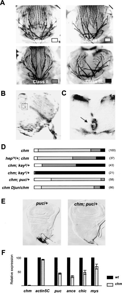
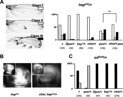
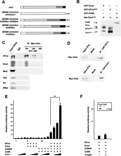
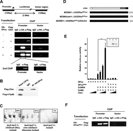
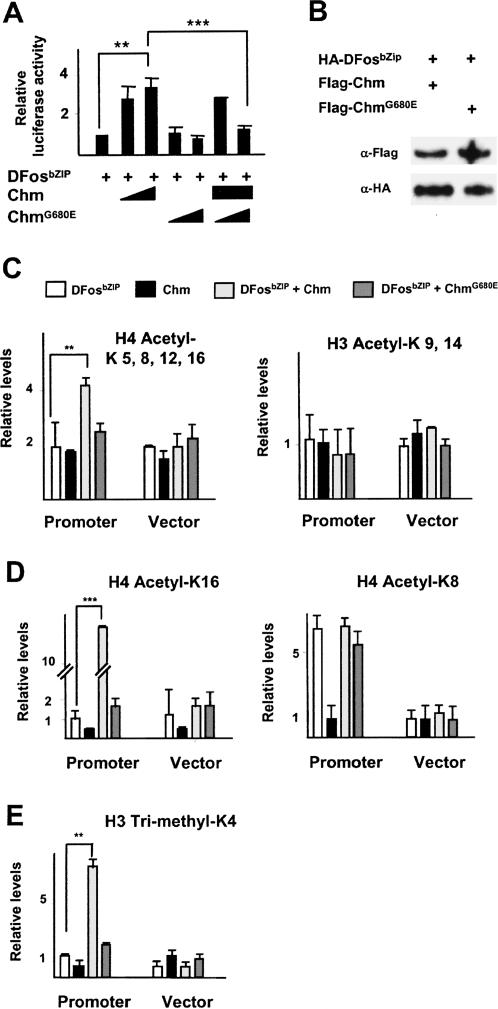
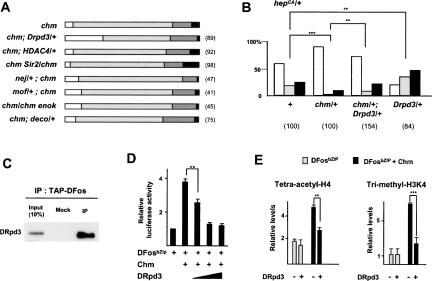
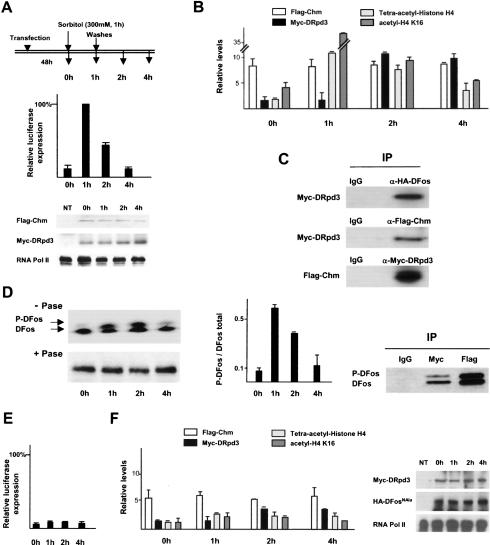
Gene regulation by AP-1 transcription factors in response to Jun N-terminal kinase (JNK) signaling controls essential cellular processes during development and in pathological situations. Here, we report genetic and molecular evidence that the histone acetyltransferase (HAT) Chameau and the histone deacetylase DRpd3 act as antagonistic cofactors of DJun and DFos to modulate JNK-dependent transcription during thorax metamorphosis and JNK-induced apoptosis in Drosophila. We demonstrate in cultured cells that DFos phosphorylation mediated by JNK signaling plays a central role in coordinating the dynamics of Chameau and DRpd3 recruitment and function at AP-1-responsive promoters. Activating the pathway stimulates the HAT function of Chameau, promoting histone H4 acetylation and target gene transcription. Conversely, in response to JNK signaling inactivation, DRpd3 is recruited and suppresses histone acetylation and transcription. This study establishes a direct link among JNK signaling, DFos phosphorylation, chromatin modification, and AP-1-dependent transcription and its importance in a developing organism.


Figures

Figure 1.
Figure 1.
Chm enhances JNK signaling during thorax closure. (A) Variable penetrance of chm thoracic cleft phenotype. (B) chm expression in peripodial cells as revealed by β-Gal activity in the large peripodial nuclei of wing discs from the chm7lacZ enhancer trap line. The inset shows the region of the disc that has been magnified. (C) chm expression at sites of imaginal disc fusion revealed by in situ hybridization on dissected pupae 8 h APF. Arrow indicates the dorsal midline. (D) Genetic interactions between chm and JNK pathway components encoding genes hep, kay, puc, and Djun, as revealed by changes in the strength of the cleft phenotype. For the color code representing the different phenotypic classes refer to A. The numbers of examined pupae are shown in parentheses. (E) puc expression in wild-type (left) and chm mutant (right) discs, revealed by β-Gal activity in the enhancer trap line pucE69. (F) Transcription levels of chm; of actin5C, which is not an AP-1 target; and of the target genes puc, mys, ance, and chic in wild-type and chm wing discs 1 h APF.
Figure 2.
Figure 2.
chm is required for JNK-induced apoptosis. (A) Variable severity of the notched wing phenotype induced by hepCA (left, arrows show wing notches) and changes induced in the relative abundance of the phenotypic classes by heterozygosis for Djun, kay, puc, chm, and chm plus puc (right). (B) Acridine orange staining of hepCA and chm; hepCA/+ wing discs. (C) Changes in the sdGal4/+ notched wing phenotype induced by puc, Djun, kay, or chm heterozygosis. The numbers of examined wings are shown in parentheses.
Figure 3.
Figure 3.
Chm acts as a nuclear coactivator of DJun and DFos. (A) Rescue of chm thoracic cleft phenotype by Chm, DFos, DJun, or DJun plus DFos expression in the MZ980 domain. Refer to Figure 1A for the color code of phenotypic classes. (B) GST pull-down assays. Binding of His-ChmCter to GST fusions of DJun, DFosN-ter or DJNK was revealed by Western blot probed by anti-His and anti-GST antibodies, and secondary antibodies coupled to peroxydase (brown) and alkaline phosphatase (blue). (C) Immunoprecipitation of nuclear extracts from chm; 69BGal4/UASMyc-chm third instar larvae using IgG or anti-Myc. Immunoprecipitates were washed by increasing salt concentration and analyzed by Western blot using antibodies to DFos, DJun, Mod, En, Ubx, and DMyc as a negative control. (D) Western blots probed with anti-Myc of immunoprecipitates obtained by anti-TAP from nuclear extracts of larvae ubiquitously expressing Myc-Chm and TAP-DFos or Myc-Chm and TAP-DJun, or of wild-type larvae (Mock). (E) Luciferase assays of HEK293 cells transfected with the reporter construct and combinations of expression vectors for DJNKK, DJNK, DFos, DJun, and Chm (50, 100, and 250 ng/well). Histograms represent luciferase activity relative to a value of 1 for transfection with reporter alone (error bars show standard deviations, five independent assays). (F) Luciferase assays with DJNKK, DJNK, DFos, DJun, and hTip60 (250 ng/well). Reference value of 1 in the absence of hTip60; four independent assays. (Inset) Western blot probed with anti-Flag of cell extracts transfected with a Flag-hTip60-producing or empty vector.
Figure 4.
Figure 4.
Cooperation between Chm and DFos in transcriptional activation. (A) ChIP with IgG, anti-HA, and anti-Flag of chromatin extracts from HEK293 cells transfected with vectors producing Flag-Chm, HA-DFos, or both. (Top) Occupancy was assayed by real-time PCR using primers for regions (shown in black) encompassing AP-1-binding sites (left) or a vector region (right). (Bottom) Second ChIP with anti-Flag from chromatin complexes precipitated by anti-HA. (B) Western blots probed with anti-Flag of immunoprecipitates obtained by anti-HA or IgG from nuclear extracts of HEK293 cells transfected with vectors producing HA-DFos and Flag-Chm (top) or HA-DFos and Flag-hTip60 (bottom). (C) Magnified views of larval epidermis stained for β-galactosidase upon lexA–lacZ reporter induction by LexA–DFos chimera in an otherwise wild-type (left) or chm mutant (middle) background, and of lexA–lacZ animals as a control (right). Levels of β-galactosidase expression are given as the percentage of the value obtained for lexA–lacZ/+; UASLexA–Dfos; hsGal4 larvae. (D) Lack of genetic interactions of chm with the DfosNAla variant during thorax closure. Refer to Figure 1A for the color code of phenotypic classes. (E) Luciferase assays of HEK293 cells transfected with DFos and Chm (50, 100, and 250 ng/well) in the presence or absence of JNK and DJNKK, and with DFosNAla and Chm in the presence of JNK and DJNKK. Reference value of 1 for transfection with reporter alone, four independent assays. (Inset) Western blot, probed with anti-DFos antibody, of cell extracts transfected with an empty vector (lane 1) and with vectors producing DFosNAla (lane 2) or DFos (lane 3). (F) ChIP with IgG, anti-HA, and anti-Flag of chromatin extracts from HEK293 cells transfected with vectors producing Flag-Chm and HA-DFosNAla. Occupancy was assayed by real-time PCR using primers for promoter and vector regions.
Figure 5.
Figure 5.
Chm promotes H4 acetylation at AP-1-responsive sequence. (A) Luciferase assays of HEK293 cells transfected with DFosbZIP, Chm, and/or ChmG680E (100 or 250 ng/well). Reference value of 1 for transfection with DFosbZIP alone; three independent assays. (B) Western blots, probed with antibody to Flag and HA tags, of chromatin preparations from cells transfected with vectors producing HA-DFosbZIP and Flag-Chm or Flag-ChmG680E. (CE) ChIP of chromatin extracts from HEK293 cells transfected with DFosbZIP, Chm, DFosbZIP and Chm, or DFosbZIP and ChmG680E. Immunoprecipitations were performed by antibodies to tetra-acetylated H4 (C, left), acetylated H3K9/K14 (C, right), acetylated H4K16 (D, left), acetylated H4K8 (D, right), and trimethylated H3K4 (E). Data of real-time PCR are given as relative levels compared with cells transfected with reporter alone (average of four independent assays).
Figure 6.
Figure 6.
DRpd3 interacts with DFos and counteracts Chm function. (A) Genetic interactions of chm with HDAC- and HAT-encoding genes during thorax closure, revealed by the strength of the thoracic cleft phenotype. The numbers of examined pupae are given in parentheses. Refer to Figure 1A for the color code of phenotypic classes. (B) Changes in the distribution of the wing phenotype classes associated with hepCA (see Fig. 2A) by heterozygosis for Drpd304556 allele and suppression of the rescuing effect of chm heterozygosis. Refer to Figure 2A for the color code of phenotypic classes. (C) Western blots, revealed by anti-DRpd3, of immunoprecipitates obtained by anti-TAP from nuclear extracts of wild-type (Mock) and armGal4 UASTAP-Dfos/UASMyc-chm third instar larvae. (D) Luciferase assays of HEK293 cells transfected with DFosbZIP, Chm (250 ng/well), and increasing amounts of DRpd3 (50, 100, or 250 ng/well). Reference value of 1 for cells transfected with DFosbZIP alone; four independent assays. (E) ChIP of chromatin extracts from HEK293 cells transfected with DFosbZIP or DFosbZIP and Chm, in the presence or absence of DRpd3. Immunoprecipitations were performed with antibodies to tetra-acetylated H4 (left) and to trimethylated H3K4 (right). Data are represented as a relative levels compared with transfection with the reporter alone (average of three independent assays).
Figure 7.
Figure 7.
Chm and DRpd3 recruitment and function upon JNK signaling activation and deactivation. (AC) Compilation of data obtained from RT–PCR, ChIP, and Western blot analyses during JNK signaling activation and inhibition upon sorbitol addition (300 mM for 1 h) and removal in HEK293 cells transfected with HA-DFos, Flag-Chm, and Myc-DRpd3. (A, top) At each time point described, total RNA was harvested for RT–PCR to access luciferase transcription, and chromatin extract was prepared for ChIP analyses. (Middle) luciferase transcription. (Bottom) Control Western blots of cell extracts for Chm and DRpd3 expression, and RNA pol II as a loading control. (NT) Nontransfected cells. (B) ChIP with anti-Flag, anti-Myc, anti-tetra-acetylated H4, and anti-acetyled H4K16 antibodies. RT–PCR data are normalized relative to GAPDH expression. ChIP data are depicted as relative levels compared with transfection with the reporter alone (average of three independent assays). (C) Western blots, probed for proteins indicated to the left, of proteins associated with chromatin fragments extracted 1 h after sorbitol removal and immunoprecipitated by antibodies to Flag (Chm), Myc (DRpd3), or HA (DFos). (D, left) Phosphorylation status of chromatin-bound HA-DFos was assessed by Western blot of chromatin samples probed by anti-HA. +Pase and –Pase refer to chromatin samples digested or not with calf intestinal alkaline phosphatase. (Middle) Time course upon sorbitol addition and removal of P-DFos/total DFos ratio obtained by phosphoimager gel scan. (Right) Western blot for DFos and P-DFos of chromatin fragments extracted 1 h after sorbitol removal and immunoprecipitated by Myc-DRpd3 and Flag-Chm. (E,F) Compilation of data obtained from similar experiments as in A and B, but using HA-DFosNAla instead of HA-DFos (average of four independent assays). (Right) Western blots of cell extracts for DRpd3, DFosNAla, and RNA pol II.

Similar articles

Cited by

References

    1. Adachi-Yamada T., Fujimura-Kamada, K., Nishida, Y., and Matsumoto, K. 1999. Distortion of proximodistal information causes JNK-dependent apoptosis in Drosophila wing. Nature 400: 166–169. - PubMed
    1. Agalioti T., Chen, G., and Thanos, D. 2002. Deciphering the transcriptional histone acetylation code for a human gene. Cell 111: 381–392. - PubMed
    1. Aggarwal B.D. and Calvi, B.R. 2004. Chromatin regulates origin activity in Drosophila follicle cells. Nature 430: 372–376. - PubMed
    1. Agnes F., Suzanne, M., and Noselli, S. 1999. The Drosophila JNK pathway controls the morphogenesis of imaginal discs during metamorphosis. Development 126: 5453–5462. - PubMed
    1. Akhtar A. and Becker, P.B. 2000. Activation of transcription through histone H4 acetylation by MOF, an acetyltransferase essential for dosage compensation in Drosophila. Mol. Cell 5: 367–375. - PubMed

Publication types

MeSH terms