

doi: 10.1021/jm050892j.
Anthrax lethal factor protease inhibitors: synthesis, SAR, and structure-based 3D QSAR studies
Affiliations
- PMID: 16392787
- PMCID: PMC3164827
- DOI: 10.1021/jm050892j
Item in Clipboard
Anthrax lethal factor protease inhibitors: synthesis, SAR, and structure-based 3D QSAR studies
J Med Chem.
.
Abstract
We have recently identified a series of compounds that efficiently inhibit anthrax lethal factor (LF) metallo-protease. Here we present further structure-activity relationship and CoMFA (comparative molecular field analysis) studies on newly derived inhibitors. The obtained 3D QSAR model was subsequently compared with the X-ray structure of the complex between LF and a representative compound. Our studies form the basis for the rational design of additional compounds with improved activity and selectivity.
Figures
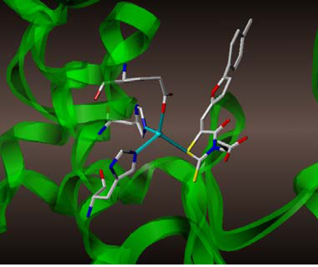
Detail of the X-ray structure of compound 1 in complex with LF (PDB_ID 1ZXV). Side chains of Zn2+ coordinating amino-acids are displayed.
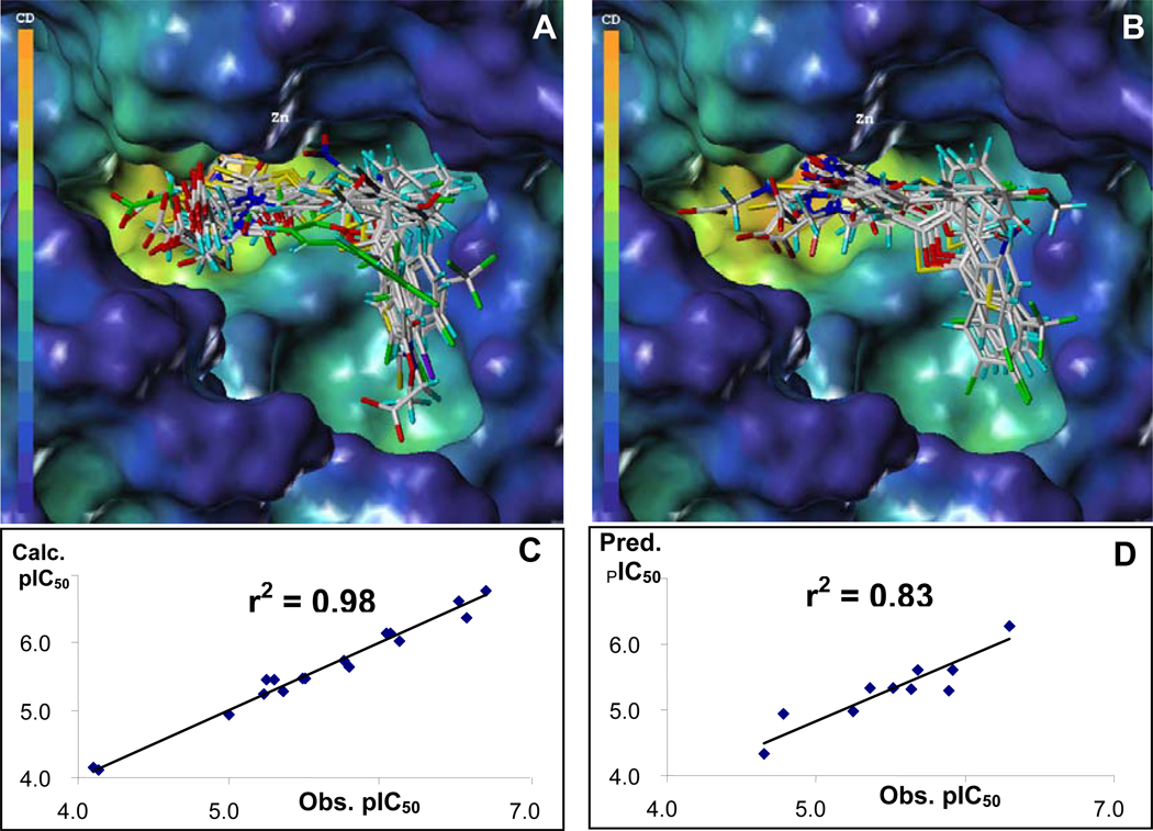
Superimpositions of docked conformers used for CoMFA studies. In (A), the structures of the compounds for the training set are displayed, with the compound highlighted in green being compound 1 (whose coordinates are from the PDB_ID 1ZXV). In (B), the aligned structures for the compounds in the test sets are displayed. (C) Calculated versus observed pIC50 values against LF for the compounds in the training set (q2 = 0.51, r2 = 0.98, # components = 4, # compounds = 17). (D) Predicted versus observed pIC50 values against LF for the 10 compounds in the test set.
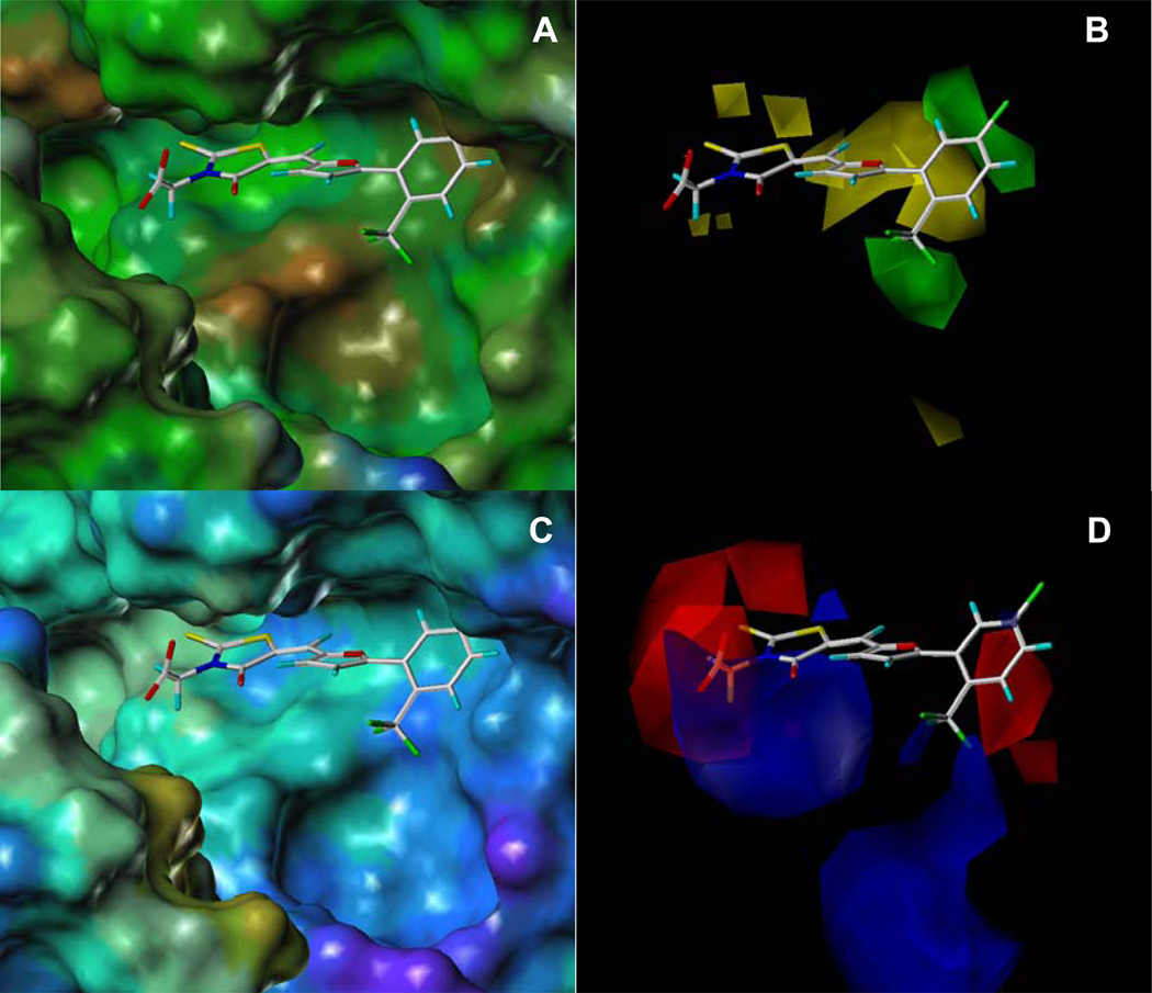
Comparison of (A) hydrophobic and hydrophilic potential molecular surface (MOLCAD) of the substrate binding site of LF in complex with compound 8 with (B) CoMFA contour plots of steric field contributions. Comparison of the (C) electrostatic potential molecular surfaces (MOLCAD) with (D) CoMFA contour plots of electrostatic field contributions. In (A), the hydrophobic and hydrophilic areas are displayed in brown and blue, respectively, while green surfaces represent an intermediate hydrophobicity. In (B), green contours indicate the regions where the addition of bulky groups may increase activity and yellow contours indicate the regions where the addition of bulky groups may decrease activity. In (C), positive and negative areas are displayed in red and blue, respectively, while cyan surfaces represent neutral areas. The color code follows the definitions of MOLCAD. In (D), blue contours indicate regions where less electronegative groups may increase activity. Red contours indicate regions where more electronegative groups may increase activity.
Similar articles
-
Guanidinylated 2,5-dideoxystreptamine derivatives as anthrax lethal factor inhibitors.Bioorg Med Chem Lett. 2006 Mar 15;16(6):1527-31. doi: 10.1016/j.bmcl.2005.12.038. Epub 2006 Jan 4. Bioorg Med Chem Lett. 2006. PMID: 16386899
-
QSAR and molecular docking studies of lethal factor protease inhibitors against Bacillus anthracis.SAR QSAR Environ Res. 2019 Oct;30(10):715-731. doi: 10.1080/1062936X.2019.1658219. Epub 2019 Sep 26. SAR QSAR Environ Res. 2019. PMID: 31556709
-
Rhodanine derivatives as selective protease inhibitors against bacterial toxins.Chem Biol Drug Des. 2008 Feb;71(2):131-9. doi: 10.1111/j.1747-0285.2007.00617.x. Epub 2008 Jan 19. Chem Biol Drug Des. 2008. PMID: 18221310
-
Discovery and development of anthrax lethal factor metalloproteinase inhibitors.Curr Pharm Biotechnol. 2008 Feb;9(1):24-33. doi: 10.2174/138920108783497604. Curr Pharm Biotechnol. 2008. PMID: 18289054 Review.
-
Quickening the pace of anthrax research: three advances point towards possible therapies.Trends Microbiol. 2002 Feb;10(2):58-62. doi: 10.1016/s0966-842x(01)02294-6. Trends Microbiol. 2002. PMID: 11827799 Review.
Cited by
-
Chelator fragment libraries for targeting metalloproteinases.ChemMedChem. 2010 Feb 1;5(2):195-9. doi: 10.1002/cmdc.200900516. ChemMedChem. 2010. PMID: 20058293 Free PMC article.
-
Cationic polyamines inhibit anthrax lethal factor protease.BMC Pharmacol. 2006 Jun 8;6:8. doi: 10.1186/1471-2210-6-8. BMC Pharmacol. 2006. PMID: 16762077 Free PMC article.
-
A high-throughput screening approach to anthrax lethal factor inhibition.Bioorg Chem. 2007 Aug;35(4):306-12. doi: 10.1016/j.bioorg.2006.12.005. Epub 2007 Feb 22. Bioorg Chem. 2007. PMID: 17320146 Free PMC article.
-
Identification of novel non-hydroxamate anthrax toxin lethal factor inhibitors by topomeric searching, docking and scoring, and in vitro screening.J Chem Inf Model. 2009 Dec;49(12):2726-34. doi: 10.1021/ci900186w. J Chem Inf Model. 2009. PMID: 19928768 Free PMC article.
-
3-Phenyl-2-thioxo-1,3-thia-zolidin-4-one.Acta Crystallogr Sect E Struct Rep Online. 2008 Sep 24;64(Pt 10):o1998. doi: 10.1107/S1600536808030079. Acta Crystallogr Sect E Struct Rep Online. 2008. PMID: 21201196 Free PMC article.
References
-
- Smith H, Keppie J. Observations on experimental anthrax: demonstration of a specific lethal factor produced in vivo by Bacillus anthracis. Nature. 1954;173:869–870. - PubMed
-
- Hanna P. Anthrax pathogenesis and host response. Curr. Topics Microbiol. Immunol. 1998;225:13–35. - PubMed
-
- Stubbs MT. Anthrax X-rayed: new opportunities for bio-defense. TRENDS Pharmacol Sci. 2002;23:539–541. - PubMed
-
- Bradley KA, Mogridge J, Mourez M, Collier RJ, Young JA. Identification of the cellular receptor for anthrax toxin. Nature. 2001;414:225–229. - PubMed
Publication types
MeSH terms
Substances
Grants and funding
LinkOut - more resources
Full Text Sources
Other Literature Sources
Chemical Information
Miscellaneous
