. 2006 Mar;17(3):1218-27.
doi: 10.1091/mbc.e05-06-0533. Epub 2006 Jan 4.

Distinct ceramide synthases regulate polarized growth in the filamentous fungus Aspergillus nidulans

Affiliations

Distinct ceramide synthases regulate polarized growth in the filamentous fungus Aspergillus nidulans

Shaojie Li et al. Mol Biol Cell. 2006 Mar.

Abstract

In filamentous fungi, the stabilization of a polarity axis is likely to be a pivotal event underlying the emergence of a germ tube from a germinating spore. Recent results implicate the polarisome in this process and also suggest that it requires localized membrane organization. Here, we employ a chemical genetic approach to demonstrate that ceramide synthesis is necessary for the formation of a stable polarity axis in the model fungus Aspergillus nidulans. We demonstrate that a novel compound (HSAF) produced by a bacterial biocontrol agent disrupts polarized growth and leads to loss of membrane organization and formin localization at hyphal tips. We show that BarA, a putative acyl-CoA-dependent ceramide synthase that is unique to filamentous fungi mediates the effects of HSAF. Moreover, A. nidulans possesses a second likely ceramide synthase that is essential and also regulates hyphal morphogenesis. Our results suggest that filamentous fungi possess distinct pools of ceramide that make independent contributions to polarized hyphal growth, perhaps through the formation of specialized lipid microdomains that regulate organization of the cytoskeleton.


Figures
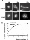
Figure 1.
Figure 1.
HSAF inhibits germ tube emergence. Conidia from wild-type strain A28 were germinated in YGV (A; top row) or YGV + 20 μg/ml HSAF (A; bottom row) for the indicated times and then stained with Hoechst 33258 and Calcofluor. Results from the time course are presented graphically in B. Bar, 3 μm.
Figure 2.
Figure 2.
Morphological effects of HSAF on A. nidulans. Wild-type hyphae (strain A28) grown on YGV were shifted to YGV (control) or YGV + 20 μg/ml HASF for the indicated time. Cell walls and sterols were visualized by staining with Calcofluor and filipin, respectively. SepA was observed in hyphae expressing SepA-GFP (strain AKS70), and microfilaments were detected using TpmA-GFP (strain ACP115). Bar, 3 μm.
Figure 3.
Figure 3.
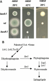
Mutants with altered responses to HSAF. (A) HSAF and temperature sensitivity of barA1, basA1, and wild-type strains. Conidia were inoculated onto MAG or MAG + 50 μg/ml HSAF plates and incubated at the indicated temperature for 3 d. (B) A simplified scheme of the Acyl-CoA-dependent sphingolipid synthesis pathway in yeast.
Figure 4.
Figure 4.
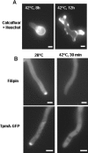
Functional characterization of BarA. (A) Wild-type (wt) and barA 1 conidia were germinated in YGV at 42°C for 8 or 12 h and then stained with Hoechst 33258 and Calcofluor. (B) Wild-type and barA1 hyphae grown in YGV were shifted to fresh YGV at either 28° or 42°C. After 30 min, hyphae were stained with filipin to observe sterols. Microfilaments were detected in wild-type and barA1 hyphae expressing TpmA-GFP. Bar, 3 μm.
Figure 5.
Figure 5.
Functional characterization of BasA. (A) basA1 conidia were germinated in YGV at 42°C for 8 or 12 h and then stained with Heochst 33258 and Calcofluor. (B) basA1 hyphae were shifted into fresh YGV medium and incubated at 28 or 42°C for 30 min. Sterols were visualized by staining with filipin. Microfilaments were detected by localizing TpmA-GFP. Bar, 3 μm.
Figure 6.
Figure 6.
Fungal Lag1-related acyl-CoA-dependent ceramide synthases. Predicted coding regions of Lag1 homologues were aligned using ClustalW (MacVector v7.0). The tree was constructed using the neighbor joining method with bootstrap support (1000 repetitions) and Poisson correction. All sequences are designated according to their annotation format or known protein name. A. nidulans (An), N. crassa (Nc), and F. graminearum (Fg) sequences were obtained from the Fungal Genome Initiative (http://www.broad.mit.edu/annotation/fungi/fgi/). F. verticillioides (Fum17 and Fum18), S. pombe (Sp), Ashbya gossypii, (Ag), S. cerevisiae (Sc), and human (Hs) sequences were obtained from NCBI. The blue line indicates BarA homologues, and the red line indicates LagA homologues.
Figure 7.
Figure 7.
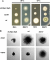
Functional characterization of LagA. (A) Comparison of growth defects observed in barA1 and lagA108 mutants. Conidiospores from strains ASL10 (alcA(p)::lagA), UV13 (barA1) and A28 (wild type) were inoculated on alcA(p)-inducing (MNVTF) or -repressing (MAG) media and incubated at the indicated temperatures for 3 d. (B) The alcA(p)::lagA mutant is sensitive to HSAF. Conidiospores with the indicated genotypes were inoculated onto MAG + 50 μg/ml HSAF plates and incubated for 24 h. Images were acquired using an Olympus SZX12 dissecting microscope (Lake Success, NY).
Figure 8.
Figure 8.
Phenotype of barA lagA double mutants. (A and B) Wild-type (A28), barA1 (UV13), alcA(p)::GFP::lagA (ASL10), and barA1 alcA(p)::GFP::lagA (DRG1; ΔΔ) conidia were germinated in alcA(p) repressing YGV at 42°C for 12 (A) or 16 h (E). (C) Conidiospores from strains UV13, ASL10, and DRG1 were inoculated on alcA(p)-inducing (MNVTF) or -repressing (MAG) media and incubated at 28°C for 3 d. Bar, 10 μm.
Figure 9.
Figure 9.
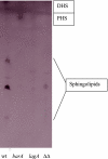
Sphingolipid levels are reduced in barA and lagA mutants. The de novo biosynthesis of sphingolipids was examined in hyphae labeled with [4,5-[3H]DHS. Lipids were extracted and analyzed by TLC as described in the Materials and Methods. The following strains were used; GR5 (wt), UV13 (barA1), ASL10 (alcA(p):: lagA), and DRG1 (ΔΔ). The migration of sphingolipids, PHS, and DHS is based on known standards.
Figure 10.
Figure 10.
Schematic representation of ceramide synthesis pathways in A. nidulans. BarA and LagA are proposed to direct the synthesis of distinct ceramide pools that independently regulate polarized hyphal growth. Through “X,” the hypothetical target of HSAF, BarA-dependent ceramides are implicated in the localization of SepA. The morphogenetic functions affected by the LagA-dependent pool remain unknown (see text for details). The dashed line indicates the possibility that HSAF could directly target BarA.

References

    1. Bagnat, M., and Simons, K. (2002). Cell surface polarization during yeast mating. Proc. Natl. Acad. Sci. USA 99, 14183–14188. - PMC - PubMed
    1. Bartnicki-Garcia, S. (2002). Hyphal tip growth: outstanding questions. In: Molecular Biology of Fungal Development, ed. H. D. Osiewacz, New York: Marcel Dekker, 29–58.
    1. Bartnicki-Garcia, S., and Lippman, E. (1969). Fungal morphogenesis: cell wall construction in Mucor rouxii. Science 165, 302–304. - PubMed
    1. Barz, W. P., and Walter, P. (1999). Two endoplasmic reticulum (ER) membrane proteins that facilitate ER-to-Golgi transport of glycosylphosphatidylinositol-anchored proteins. Mol. Biol. Cell 10, 1043–1059. - PMC - PubMed
    1. Bauer, Y., Knechtle, P., Wendland, J., Helfer, H., and Philippsen, P. (2004). A Ras-like GTPase is involved in hyphal growth guidance in the filamentous fungus Ashbya gossypii. Mol. Biol. Cell 15, 4622–4632. - PMC - PubMed

Publication types

MeSH terms