. 2006 Mar;17(3):1421-35.
doi: 10.1091/mbc.e05-08-0802. Epub 2006 Jan 4.

The V260I mutation in fission yeast alpha-tubulin Atb2 affects microtubule dynamics and EB1-Mal3 localization and activates the Bub1 branch of the spindle checkpoint

Affiliations

The V260I mutation in fission yeast alpha-tubulin Atb2 affects microtubule dynamics and EB1-Mal3 localization and activates the Bub1 branch of the spindle checkpoint

Kazuhide Asakawa et al. Mol Biol Cell. 2006 Mar.

Abstract

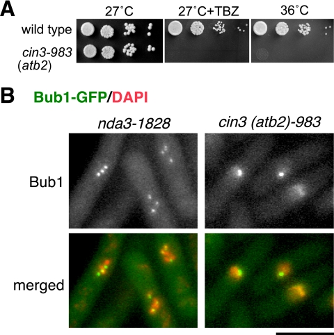
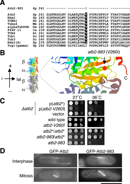
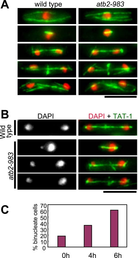
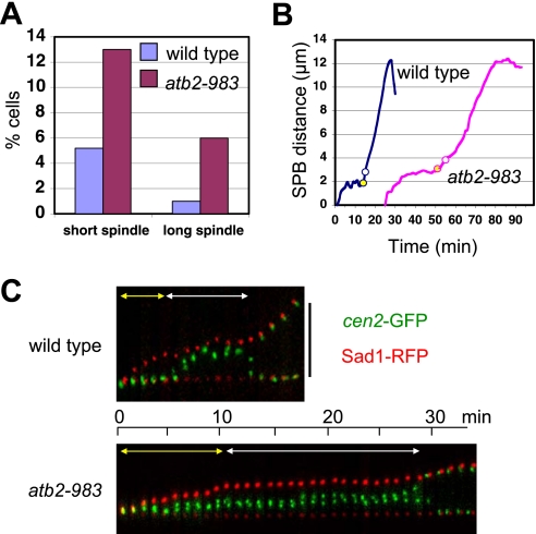
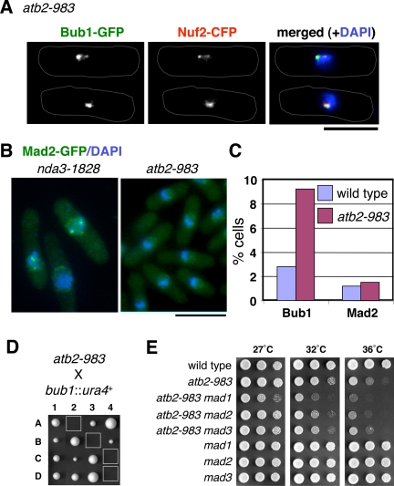
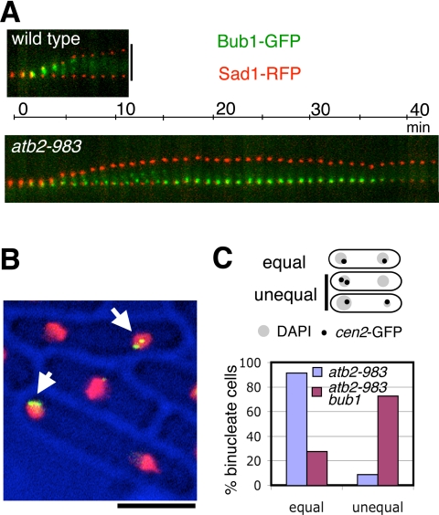
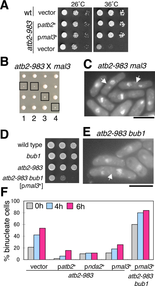
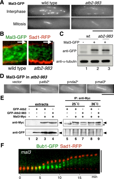
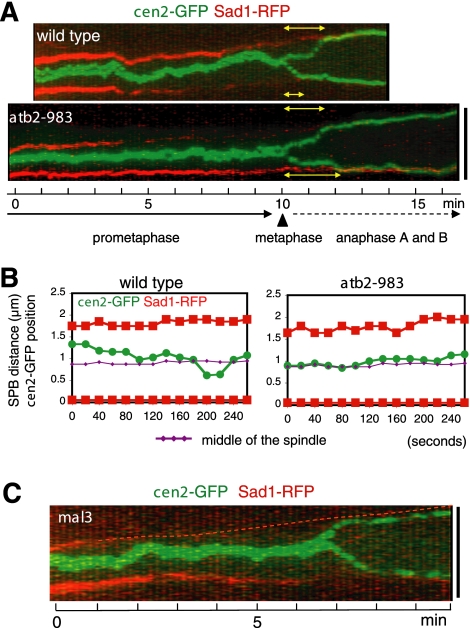
We have identified a novel temperature-sensitive mutant of fission yeast alpha-tubulin Atb2 (atb2-983) that contains a single amino acid substitution (V260I). Atb2-983 is incorporated into the microtubules, and their overall structures are not altered noticeably, but microtubule dynamics is compromised during interphase. atb2-983 displays a high rate of chromosome missegregation and is synthetically lethal with deletions in a subset of spindle checkpoint genes including bub1, bub3, and mph1, but not with mad1, mad2, and mad3. During early mitosis in this mutant, Bub1, but not Mad2, remains for a prolonged period in the kinetochores that are situated in proximity to one of the two SPBs (spindle pole bodies). High dosage mal3(+), encoding EB1 homologue, rescues atb2-983, suggesting that Mal3 function is compromised. Consistently, Mal3 localization and binding between Mal3 and Atb2-983 are impaired significantly, and a mal3 single mutant, such as atb2-983, displays prolonged Bub1 kinetochore localization. Furthermore in atb2-983 back-and-forth centromere oscillation during prometaphase is abolished. Intriguingly, this oscillation still occurs in the mal3 mutant, indicating that there is another defect independent of Mal3. These results show that microtubule dynamics is important for coordinated execution of mitotic events, in which Mal3 plays a vital role.


Figures

Figure 1.
Figure 1.
Isolation of the cin3-983 mutant. (A) Temperature and TBZ sensitivity. Wild-type (513; Table 1) or cin3 (atb2)-983 mutants (KZ94) were spotted on rich medium (5 × 10³ cells in the far-left spots for each plate and then diluted 10-fold in each subsequent spot rightward) in the presence (middle plate) or absence (left and right) of 10 μg/ml TBZ and incubated at 27°C (left and middle) or at 36°C (right) for 3 d. (B) Localization of Bub1-GFP. nda3-1828 (KZ35) or cin3 (atb2)-983 cells (ts983) were incubated at 36°C for 4 h and Bub1-GFP and DNA were visualized. The top panels show Bub1-GFP signals, whereas the bottom panels show merged pictures (Bub1-GFP in green and DAPI in orange). Bar, 10 μm.
Figure 2.
Figure 2.
The cin3-983 mutant is allelic to atb2. (A) The mutation site of Atb2-983. The valine 260 residue (white letters in black boxes) was substituted with isoleucine (red) in the atb2-983 mutant. The regions around the valine 260 in α-tubulins from various species were aligned. The corresponding regions in fission yeast β-tubulin (Nda3) and γ-tubulin (Tug1/Gtb1) are also shown. Sp, Schizosaccharomyces pombe; Hs, Homo sapiens; Mm, Mus muscalas; Dm, Drosophila melanogaster; At, Arabidopsis thaliana; Ce, Caenorhabditis elegans; and Sc, Saccharomyces cerevisiae. (B) Predicted microtubule structure. The structure of fission yeast α/β-tubulin dimers was predicted by RasMol software and aligned longitudinally based on bovine α/β-tubulin protofilament (Nogales et al., 1998). Structure is oriented with the plus end upward (+), the minus end downward (–), the inside leftward (i), and the outside rightward (lat). The location of the atb2-983 mutation was indicated by arrow with red circle in a magnified view of the interdimer interface (right). (C) The atb2-V260I mutation confers temperature sensitivity. p(atb2+), p(atb2-V260I) or vector plasmids were introduced into an atb2-deletion strain (KZ115, top). A strain containing the integrated atb2-V260I allele at its genomic locus (KZ217, middle), wild-type diploids (atb2+/atb2+, bottom) and heterozygous diploids (atb2-983/atb2+) were also tested for temperature sensitivity. Each strain was spotted on rich medium and incubated at 27 or 36°C for 3 d. (D) Incorporation of GFP-Atb2-983 into the microtubules. A strain containing wild-type GFP-atb2+ (P3-GFP-atb2) or mutant GFP-atb2-983 (KZ176) was grown at 27°C in the liquid YE5S medium, and GFP signals were recorded. Images of interphase (top) and mitosis (bottom) are shown. Bar, 10 μm.
Figure 3.
Figure 3.
The atb2-983 mutant shows unequal chromosome segregation. (A) Microtubule structure in atb2-983 cells during the cell cycle. Wild-type (513) and atb2-983 cells (KZ94) were incubated at 36°C for 4 h, fixed, and processed for immunofluorescence microscopy with anti-α-tubulin antibody (TAT-1; green) and DAPI (red). Representative images of wild-type (left) and atb2-983 cells (right) in each cell cycle stage are shown (from the top, interphase, early mitosis, late anaphase, telophase, and postmitosis). (B) Unequal chromosome segregation in atb2-983. Wild-type (top) or atb2-983 cells (bottom three panels) were processed for immunofluorescence microscopy as in described in A. Representative cells displaying chromosome missegregation in atb2-983 are shown. Bars, 10 μm. (C) Quantification of chromosome missegregation. Wild-type and atb2-983 cells were grown at 25°C and shifted to 36°C. After 4 and 6 h, aliquots were taken, fixed, stained with DAPI, and the percentage of chromosome missegregation was counted (n = 100). We did not find any chromosome missegregation in wild-type cells under this condition.
Figure 4.
Figure 4.
Mitosis is slowed down in atb2-983 cells. (A) Mitotic delay in atb2-983 cells. Wild-type (513) and atb2-983 cells (KZ94) were incubated at 36°C for 4 h, and MTs were visualized with immunofluorescence as in Figure 3. The population of cells with short (<2 μm) or long spindle (>2 μm) was counted from >300 cells. (B) Passage of mitotic phases in live atb2-983 cells. Wild-type (KZ85) and atb2-983 cells (KZ139) containing cen2-GFP and Sad1-RFP were grown, and time-lapse live analysis was performed at the room temperature (n = 10). Typical examples for the two strains were shown. The timing of anaphase A onset and completion is marked with yellow and white circles, respectively. (C) Kymograph images of the cells shown in B. Kymograph images were produced every 1-min interval. Yellow and white arrows indicate the duration of prophase and prometaphase/metaphase, respectively. Vertical bar, 5 μm.
Figure 5.
Figure 5.
Bub1-, but not Mad2-, checkpoint is activated in atb2-983 cells. (A) Kinetochore localization of Bub1 in atb2-983 cells. Characteristic mitotic atb2-983 cells that display Bub1-GFP dots (left) at the kinetochores (Nuf2-CFP; middle) are shown (KZ144; 4 h at 36°C). In the merged image (right), Bub1-GFP (green), Nuf2-CFP (red), and DAPI (blue) are shown. (B) Mad2 recruitment to the kinetochore is not enhanced by the atb2-983 mutation. Mad2-GFP and DNA were visualized in nda3-1828 cells (KZ54; left) or atb2-983 cells (KZ156; right) after a 4-h incubation at 36°C. Bars, 10 μm. (C) Quantification of Mad2-GFP and Bub1-GFP dots. The percentage of wild-type and atb2-983 mutant cells (grown at 27°C) that display Bub1 or Mad2 dots (n > 350) is shown. (D) Synthetic lethality of an atb2-983bub1 strain. Tetrads from genetic cross between an atb2-983 strain (KZ102) and bub1::ura4+ strain (393) were dissected and incubated at 27°C for 4 d. Segregants corresponding to atb2-983bub1::ura4+ double mutants (open square) failed to form colonies. (E) Growth properties of atb2-983mad double mutants. Strains with indicated genotypes were spotted on rich medium (5 × 10⁴ cells in the far-left spots for each plate and then diluted 10-fold in each subsequent spot rightward) and incubated at 27°C (left), 32°C (middle), or 36°C (right) for 3 d.
Figure 6.
Figure 6.
The Bub1 spindle checkpoint prevents unequal chromosome segregation in atb2-983 cells. (A) Prolonged localization of Bub1 blob during mitosis. Time-lapse images of Bub1-GFP and Sad1-RFP localization during mitosis were recorded and converted to a kymograph every 1-min interval (top, wild type, KZ181; and bottom, atb2-983, KZ182). Vertical bar, 5 μm. (B) Unequal chromosome segregation phenotype of atb2-983bub1 cells. atb2-983bub1 cells containing cen2-GFP were kept viable by expressing episomal atb2+, starved for nitrogen for 12 h, and released into the rich medium (26°C) to induce plasmid loss. At 8 h after release, patterns of chromosome segregation were observed with cen2-GFP (green) and DAPI staining (red). Representative images displaying 2:0 distribution of cen2-GFP (marked with arrows) are shown. Bar, 10 μm. (C) The percentage of unequal chromosome segregation in atb2-983 and atb2-983bub1 cells.
Figure 7.
Figure 7.
Suppression of atb2-983 by multicopy mal3+ gene encoding the EB1 homolog. (A) Suppression of the ts atb2-983 mutation by multicopy plasmids containing mal3+. wt, wild-type cells. (B) Genetic interaction between atb2-983 and mal3. Tetrads from genetic cross between an atb2-983 strain (KZ137) and a mal3-deleted strain (YP350) were dissected and incubated at 27°C for 4 d. Segregants corresponding to atb2-983mal3 double mutants are marked with open squares. (C) Chromosome missegregation of atb2-983mal3 cells. The atb2-983mal3 double mutant (KZ141) was grown at 27°C and stained with DAPI. Cells displaying chromosome missegregation are pointed by arrows. (D) Suppression of the lethality of atb2-983bub1 by a high dosage of mal3+. atb2-983bub1 (pmal3+-LEU2) (KZ135) was obtained from the cross between an atb2-983 strain carrying plasmids containing mal3+ (pmal3+-LEU2) and a bub1::ura4+ strain. (E) Chromosome missegregation of atb2-983bub1 carrying multicopy mal3+. Bar, 10 μm. (F) Quantification of chromosome missegregation. Indicated strains were grown in minimal liquid media lacking leucine at 27°C and shifted to 36°C. At 0-, 4-, and 6-h time points, aliquots were taken and stained with DAPI. The percentage of binucleate cells displaying chromosome missegregation was quantified (n = 100).
Figure 8.
Figure 8.
Mal3 loading onto the spindle and binding to Atb2–983 are reduced in the atb2-983 mutant. (A) Reduced Mal3 localization. Wild-type (MA145) and ab2-983 cells containing Mal3-GFP (KZ165) were grown at 27°C and GFP signals were taken. (B) Time-lapse analysis of mitotic Mal3-GFP. The live signals of mitotic Mal3-GFP in wild-type (KZ194; left) or atb2-983 (KZ195; right) cells were obtained and converted to kymograph pictures (Mal3-GFP in green and Sad1-RFP in red). The length of the white arrow and the black bar represents 1 min and 2 μm, respectively. (C) Protein levels of Mal3 in atb2-983. Extracts were prepared from exponentially growing wild-type (lane 2) or atb2-983 cells containing Mal3-GFP (lane 3) and immunoblotted with indicated antibodies. An untagged wild-type strain (lane 1) was used as a control. (D) Suppression of compromised spindle localization of Mal3. atb2-983 mutants carrying various multicopy plasmids (an empty vector, atb2+, nda2+, or mal3+) were grown at 27°C and shifted to 36°C for 4 h, and Mal3-GFP signals were taken. Representative Mal3-GFP signals localizing to the spindle are shown (n = 20). The exposure time of these pictures are the same (2 s). (E) Immunoprecipitation between GFP-Atb2 (-983) and Mal3-Myc. Wild-type (KZ214; lanes 3, 6, and 8) or atb2-983 mutants containing GFP-Atb2 (-983) and Mal3-GFP (KZ216; lanes 4, 7, and 9) were grown at 25°C (lanes 2, 3, 6, and 7) and shifted to 36°C (lanes 8 and 9). Total cell extracts were prepared at 0 (lanes 3 and 4, 10 mg of extracts was loaded) and 4 h (lanes 5–8), and immunoprecipitation was performed using monoclonal anti-Myc antibody as a primary antibody. Immunoprecipitates were run on SDS-PAGE, and immunoblotting was performed with polyclonal anti-Myc antibody (top) or monoclonal anti-GFP antibody (bottom). As a control, immunoprecipitation was performed from extracts prepared from a strain containing only GFP-Atb2 (lanes 2 and 5). Extracts prepared from an untagged wild-type strain was also run as another control (lane 1). Precipitated bands were marked with arrowheads, and nonspecific bands, probably corresponding to IgG heavy chains, were shown with asterisk. (F) Prolonged localization of Bub1 blob during early mitosis in mal3 cells. Time-lapse images (1-min intervals) of Bub1-GFP and Sad1-RFP localization during mitosis were recorded and converted to a kymograph. For patterns of wild-type and atb2-983 cells, see Figure 6A. Vertical bars, 2 μm.
Figure 9.
Figure 9.
Lack of sister centromere oscillation in the atb2-983 mutation. (A) Visualization of sister centromere movement in live cells. Kymograph images at high resolution (10-s intervals) are shown (cen2-GFP in green and Sad1-RFP in red) in wild-type (top; n = 32) and atb2-983 cells (bottom; n = 30). cen2-GFP signals that were persistently associated with one SPB like Bub1-GFP (see Figure 6A), which probably represented syntelic attachment, were not analyzed in this assay. Yellow bidirectional arrows show the duration of anaphase A. Solid and dotted arrows indicate the duration of prometaphase and anaphase, respectively. Arrowhead indicates the timing of metaphase. (B) Prometaphase movement of cen2-GFP. The positions of cen2-GFP (green circles) are plotted relative to the two SPBs (red squares) every 20-s interval. The central position of the two SPBs is shown with purple lines (diamonds). When cen2-GFP signals seemed split, the middle position is plotted. The representative images are shown in wild-type (n = 12) and atb2-983 (n = 10). (C) Visualization of sister centromere movement in mal3 cells. Kymograph image in the mal3 mutant is constructed as described in A. Vertical bars, 5 μm.

Similar articles

Cited by

References

    1. Adachi, Y., Toda, T., and Yanagida, M. (1986). Differential expression of essential and nonessential a-tubulin genes in Schizosaccharomyces pombe. Mol. Cell. Biol. 6, 2168–2178. - PMC - PubMed
    1. Asakawa, K., and Toda, T. (2006). Cooperation of EB1-Mal3 and the Bub1 spindle checkpoint. Cell Cycle 5, 27–30. - PubMed
    1. Asakawa, K., Toya, M., Sato, M., Kanai, M., Kume, K., Goshima, T., Garcia, M. A., Hirata, D., and Toda, T. (2005). Mal3, the fission yeast EB1 homolog, cooperates with Bub1 spindle checkpoint to prevent monopolar attachment. EMBO Rep. 6, 1194–1200. - PMC - PubMed
    1. Beinhauer, J. D., Hagan, I. M., Hegemann, J. H., and Fleig, U. (1997). Mal3, the fission yeast homologue of the human APC-interacting protein EB-1 is required for microtubule integrity and the maintenance of cell form. J. Cell Biol. 139, 717–728. - PMC - PubMed
    1. Bernard, P., Hardwick, K., and Javerzat, J.-P. (1998). Fission yeast Bub1 is a mitotic centromere protein essential for the spindle checkpoint and the preservation of correct ploidy through mitosis. J. Cell Biol. 143, 1775–1787. - PMC - PubMed

Publication types

MeSH terms

LinkOut - more resources