

A method of image registration for small animal, multi-modality imaging
- PMID: 16394345
- PMCID: PMC3005360
- DOI: 10.1088/0031-9155/51/2/013
A method of image registration for small animal, multi-modality imaging
Abstract
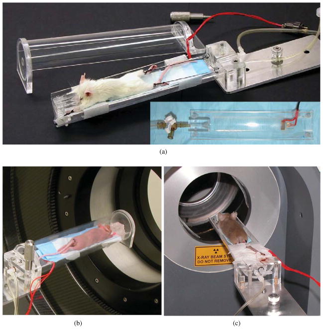
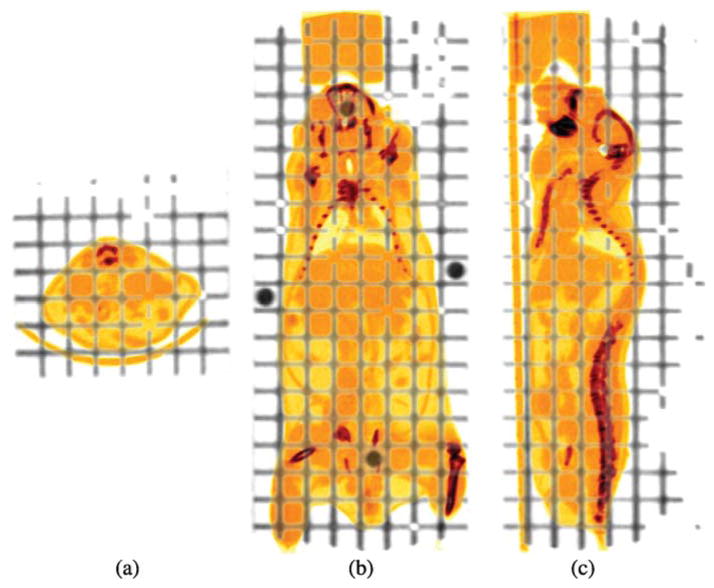
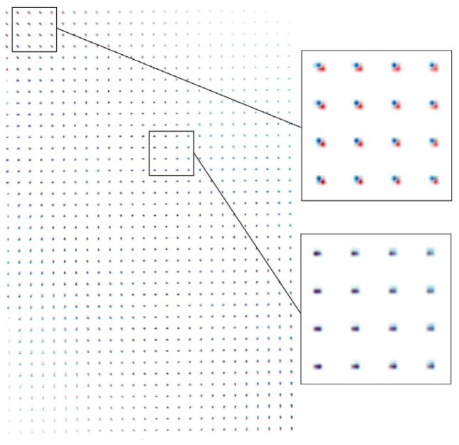
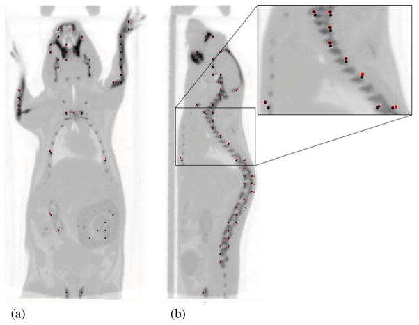
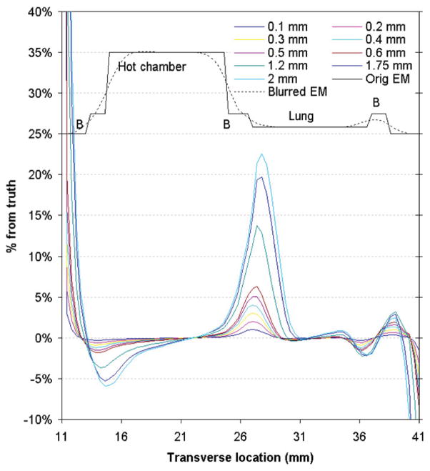
Many research institutions have a full suite of preclinical tomographic scanners to answer biomedical questions in vivo. Routine multi-modality imaging requires robust registration of images generated by various tomographs. We have implemented a hardware registration method for preclinical imaging that is similar to that used in the combined positron emission tomography (PET)/computed tomography (CT) scanners in the clinic. We designed an imaging chamber which can be rigidly and reproducibly mounted on separate microPET and microCT scanners. We have also designed a three-dimensional grid phantom with 1288 lines that is used to generate the spatial transformation matrix from software registration using a 15-parameter perspective model. The imaging chamber works in combination with the registration phantom synergistically to achieve the image registration goal. We verified that the average registration error between two imaging modalities is 0.335 mm using an in vivo mouse bone scan. This paper also estimates the impact of image misalignment on PET quantitation using attenuation corrections generated from misregistered images. Our technique is expected to produce PET quantitation errors of less than 5%. The methods presented are robust and appropriate for routine use in high throughput animal imaging facilities.
Figures
Similar articles
-
Phantom validation of coregistration of PET and CT for image-guided radiotherapy.Med Phys. 2004 May;31(5):1083-92. doi: 10.1118/1.1688041. Med Phys. 2004. PMID: 15191296
-
Impact of a multiple mice holder on quantitation of high-throughput MicroPET imaging with and without Ct attenuation correction.Mol Imaging Biol. 2013 Oct;15(5):569-75. doi: 10.1007/s11307-012-0602-y. Mol Imaging Biol. 2013. PMID: 23479323 Free PMC article.
-
Automated 3-dimensional elastic registration of whole-body PET and CT from separate or combined scanners.J Nucl Med. 2005 Sep;46(9):1488-96. J Nucl Med. 2005. PMID: 16157532 Clinical Trial.
-
Positron emission tomography/computed tomography.Semin Nucl Med. 2008 May;38(3):152-66. doi: 10.1053/j.semnuclmed.2008.01.003. Semin Nucl Med. 2008. PMID: 18396176 Review.
-
Magnetic resonance-based motion correction for positron emission tomography imaging.Semin Nucl Med. 2013 Jan;43(1):60-7. doi: 10.1053/j.semnuclmed.2012.08.007. Semin Nucl Med. 2013. PMID: 23178089 Free PMC article. Review.
Cited by
-
A method of 2D/3D registration of a statistical mouse atlas with a planar X-ray projection and an optical photo.Med Image Anal. 2013 May;17(4):401-16. doi: 10.1016/j.media.2013.02.009. Epub 2013 Mar 5. Med Image Anal. 2013. PMID: 23542374 Free PMC article.
-
Performance evaluation of PETbox: a low cost bench top preclinical PET scanner.Mol Imaging Biol. 2011 Oct;13(5):949-61. doi: 10.1007/s11307-010-0413-y. Mol Imaging Biol. 2011. PMID: 20812031 Free PMC article.
-
Importance of Attenuation Correction (AC) for Small Animal PET Imaging.Diagnostics (Basel). 2012 Oct 9;2(4):42-51. doi: 10.3390/diagnostics2040042. Diagnostics (Basel). 2012. PMID: 26859397 Free PMC article.
-
DigiWarp: a method for deformable mouse atlas warping to surface topographic data.Phys Med Biol. 2010 Oct 21;55(20):6197-214. doi: 10.1088/0031-9155/55/20/011. Epub 2010 Sep 30. Phys Med Biol. 2010. PMID: 20885019 Free PMC article.
-
Impact of tumor-specific targeting on the biodistribution and efficacy of siRNA nanoparticles measured by multimodality in vivo imaging.Proc Natl Acad Sci U S A. 2007 Sep 25;104(39):15549-54. doi: 10.1073/pnas.0707461104. Epub 2007 Sep 17. Proc Natl Acad Sci U S A. 2007. PMID: 17875985 Free PMC article.
References
-
- Chatziioannou AF, Stout DB, Silverman RW. Method and apparatus for animal positioning in imaging systems. US Provisional Patent Application. R268:53028 2004.
-
- Chow PL, Rannou FR, Chatziioannou AF. Molecular Imaging and Biology (San Diego) Vol. 4. New York: Elsevier; 2002. Attenuation correction for a 3D small animal PET tomograph, using x-ray microCT; p. S17.
-
- Defrise M. A factorization method for the 3D x-ray transform. Inverse Problems. 1995;11:983–94.
-
- Hill DLG, Batchelor PG, Holden M, Hawkes DJ. Medical image registration. Phys Med Biol. 2001;46:R1–45. - PubMed
Publication types
MeSH terms
Grants and funding
LinkOut - more resources
Full Text Sources
Other Literature Sources