Comparative Study
. 2006 Jan 25;25(2):398-408.
doi: 10.1038/sj.emboj.7600922. Epub 2006 Jan 5.

Structure of the N-terminal fragment of topoisomerase V reveals a new family of topoisomerases

Affiliations
Comparative Study

Structure of the N-terminal fragment of topoisomerase V reveals a new family of topoisomerases

Bhupesh Taneja et al. EMBO J. .

Abstract

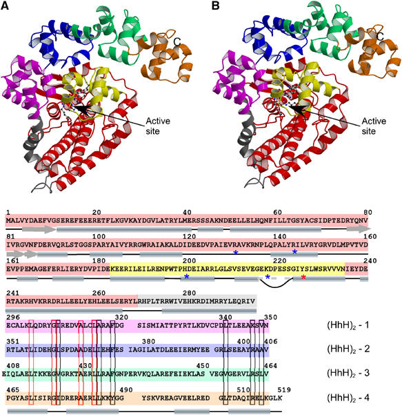
Topoisomerases are involved in controlling and maintaining the topology of DNA and are present in all kingdoms of life. Unlike all other types of topoisomerases, similar type IB enzymes have only been identified in bacteria and eukarya. The only putative type IB topoisomerase in archaea is represented by Methanopyrus kandleri topoisomerase V. Despite several common functional characteristics, topoisomerase V shows no sequence similarity to other members of the same type. The structure of the 61 kDa N-terminal fragment of topoisomerase V reveals no structural similarity to other topoisomerases. Furthermore, the structure of the active site region is different, suggesting no conservation in the cleavage and religation mechanism. Additionally, the active site is buried, indicating the need of a conformational change for activity. The presence of a topoisomerase in archaea with a unique structure suggests the evolution of a separate mechanism to alter DNA.


Figures

Figure 1
Figure 1
Overall structure of the 61 kDa amino terminal fragment of M. kandleri topoisomerase V (topo-61). (A) Stereo diagram of the structure showing the topoisomerase domain (red), the linker helix (gray) and the four (HhH)2 domains (pink, blue, green and orange). The HTH motif containing some of the active site residues is shown in yellow. The putative active site residues are shown as ball and stick. The C-terminus is marked with a C. The N-terminus resides at the beginning of the β-strand behind the HTH domain and is not marked for clarity. (B) Sequence of the topo-61 fragment showing the location of the different domains. The position of the secondary structure elements is shown by cylinders and arrows. The loop inserted in the recognition helix of the HTH motifs is shown by a curved line; the recognition helix is continuous despite this insertion. The putative active site residues are marked with an asterisk (*). The sequences of the (HhH)2 domains were aligned based on the structure. The position of the helices in the HhH motif is shown at the bottom of the alignment, but for clarity the helix linking the HhH motifs in an (HhH)2 domain is not shown. The alignment shows marked sequence similarities in the first HhH motif, but not in the second motif. Similar residues are boxed in black, identical residues are boxed in red.
Figure 2
Figure 2
The multiHhH domain is flexible, while the topoisomerase domain is rigid. Schematic diagram showing the superposition of the two monomers in the asymmetric unit of crystal Form I. The active site residues are shown in yellow. In (A) only the multiHhH domains were used for the superposition, while in (B) the topoisomerase domains were used for the superposition. The figure illustrates the variability in the multiHhH domains (Supplementary data). For clarity, in each monomer the topoisomerase domain and the multiHhH domain are shown in different shades of the same color.
Figure 3
Figure 3
Topoisomerase V active site. (A) Stereo diagram of the active site region. The location is defined by the presence of the active site tyrosine, Tyr226. Other putative residues involved in catalysis are shown, including Arg-131, Arg-144, His-200 and Lys-218. A network of interactions involving water molecules (shown in green) is present. The first (HhH)2 domain blocks access to the active site and would have to move away in order to expose the active site. The coloring of the structure corresponds to the scheme in Figure 1. (B) Mutational analysis of the putative active site residues. The amino acids were mutated to alanine and tested for relaxation activity. Five mutants were tested: Y226A, R131A, R144A, K218A and H200A. In the assay, 0.2 μg of negatively supercoiled pBR322 DNA was incubated at 75°C for 15 min with 1, 2 or 5 U of protein, respectively. Lanes 1–3, Y226A; lanes 4–6, R131A; lanes 7–9, R144A; lanes 10–12, K218A; lanes 13–15, H200A. As a control, the relaxation by 0.7 and 5 U of wild-type protein (WT) is also shown (lanes 16 and 17). Lane 18 shows plasmid DNA with no protein. The position of the supercoiled DNA is labeled as SC.
Figure 4
Figure 4
Model for a topoisomerase domain–DNA complex. (A) Electrostatic surface representation of the topoisomerase domain. The diagram shows that the topoisomerase has a large positively charged groove in one face of the protein centered around the active site. The insets correspond to 90° views of the molecule and show that there are no additional large positively charged regions in the protein. The electrostatic potential was calculated with the program APBS (Baker et al, 2001), with a dielectric constant of 80 for the solvent and 20 for the protein. The surface is colored with a blue to red gradient from +5 to −5 KbT/e. (B) The diagram shows a model of the topoisomerase domain in complex with B-DNA. The coloring scheme is the same as in Figure 1. To create the model, the linker helix and the multiHhH domain were removed. DNA was docked using the HTH motif of human Pax6-paired domain–DNA complex (Xu et al, 1999), but replacing the DNA with a canonical, straight B-DNA model. No attempts were made to prevent steric clashes. In the model, the active site tyrosine is ideally placed to interact with the phosphodiester backbone and the other putative active site residues are also in the vicinity of DNA. The model suggests a potential way for topo-61 to interact with DNA, making extensive interactions.
Figure 5
Figure 5
The 61 kDa N-terminal fragment of topoisomerase V undergoes a temperature-dependent conformational change. (A) Plot of the wavelengths of maximum emission versus temperature. A small blue shift is observed in the fluorescence emission maximum of topo-61, but not topo-44 as the temperature is increased. The blue circles and red triangles show the position of emission maximum at each temperature for topo-61 and for topo-44, respectively. A line is used as a guide to identify the two conformational states of topo-61 or the single conformational state of topo-44, respectively. The wavelength of maximum emission returns to its original value upon cooling the protein back to 25°C (green circle). The error bars correspond to the standard deviations of the measurements. (B, C) Temperature dependence of the relaxation activity of topo-44 and topo-61. Relaxation assays were performed at the different temperatures indicated. Topo-61 is not active below 50°C (lanes 1–3 in (C)), above which its activity increases with temperature (lanes 4–6 in (C)). Similarly, topo-44 shows no activity below 55°C (lanes 1–4 in (B)), above which its activity increases with temperature (lanes 5–6 in (B)). Lanes 7 and 8 in both (B) and (C) show DNA with no protein at 25 and 70°C. The conformational change in topo-61 correlates with the temperature dependence of its relaxation activity.
Figure 6
Figure 6
Topoisomerase V represents a new family of type IB topoisomerases. The figure summarizes some of the properties of the three families of type I topoisomerases. Each column shows the overall structure and the active site region of a representative of the respective family (type IA, E. coli topoisomerase III (Changela et al, 2001); type IB, human topoisomerase I (Redinbo et al, 1998); topo V, 61 kDa N-terminal fragment of topoisomerase V). Type IA enzymes form a very distinct family with no sequence, structural or mechanistic similarities to the other families. Type IB enzymes and topoisomerase V share many overall features, but there is no sequence, structure or apparent mechanistic similarities between these two families.

References

    1. Aravind L, Anantharaman V, Balaji S, Babu MM, Iyer LM (2005) The many faces of the helix–turn–helix domain: transcription regulation and beyond. FEMS Microbiol Rev 29: 231–262 - PubMed
    1. Ariyoshi M, Nishino T, Iwasaki H, Shinagawa H, Morikawa K (2000) Crystal structure of the holliday junction DNA in complex with a single RuvA tetramer. Proc Natl Acad Sci USA 97: 8257–8262 - PMC - PubMed
    1. Baker NA, Sept D, Joseph S, Holst MJ, McCammon JA (2001) Electrostatics of nanosystems: application to microtubules and the ribosome. Proc Natl Acad Sci USA 98: 10037–10041 - PMC - PubMed
    1. Belova GI, Prasad R, Kozyavkin SA, Lake JA, Wilson SH, Slesarev AI (2001) A type IB topoisomerase with DNA repair activities. Proc Natl Acad Sci USA 98: 6015–6020 - PMC - PubMed
    1. Belova GI, Prasad R, Nazimov IV, Wilson SH, Slesarev AI (2002) The domain organization and properties of individual domains of DNA topoisomerase V, a type 1B topoisomerase with DNA repair activities. J Biol Chem 277: 4959–4965 - PubMed

Publication types

Substances