. 2006 Jan;116(1):36-48.
doi: 10.1172/JCI26505.

Control of SRF binding to CArG box chromatin regulates smooth muscle gene expression in vivo

Affiliations

Control of SRF binding to CArG box chromatin regulates smooth muscle gene expression in vivo

Oliver G McDonald et al. J Clin Invest. 2006 Jan.

Abstract

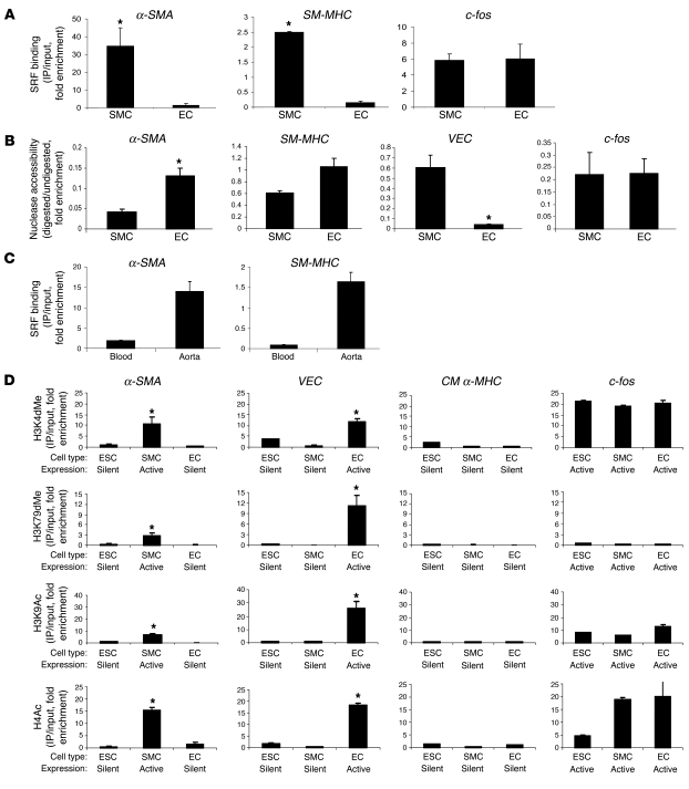
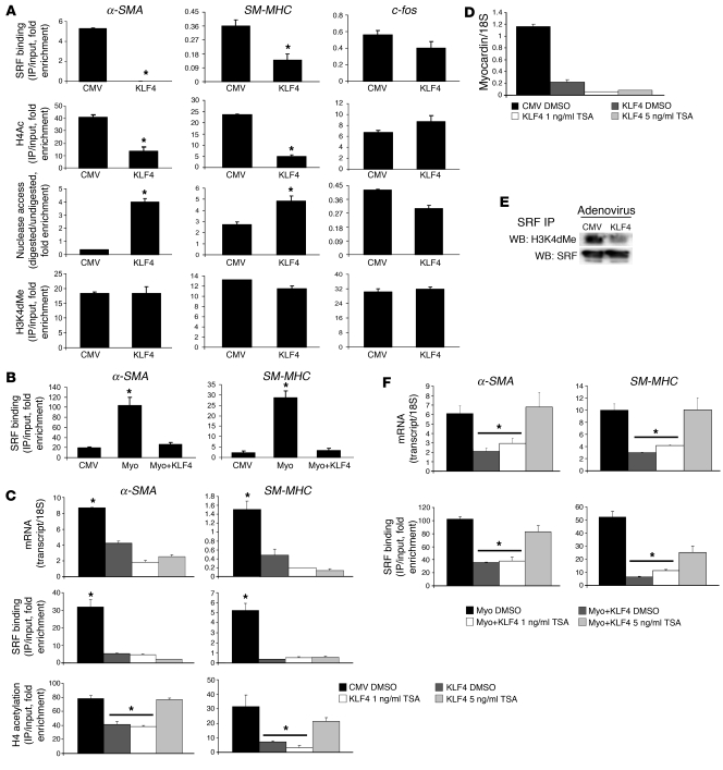
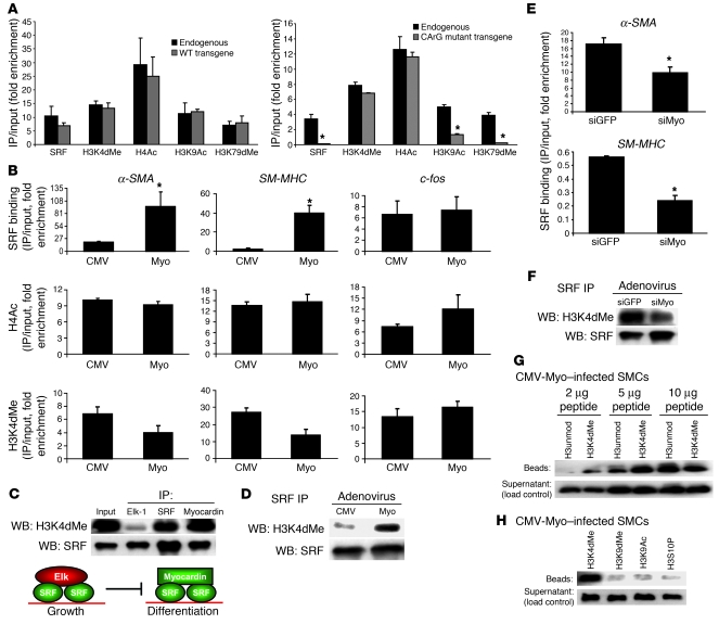
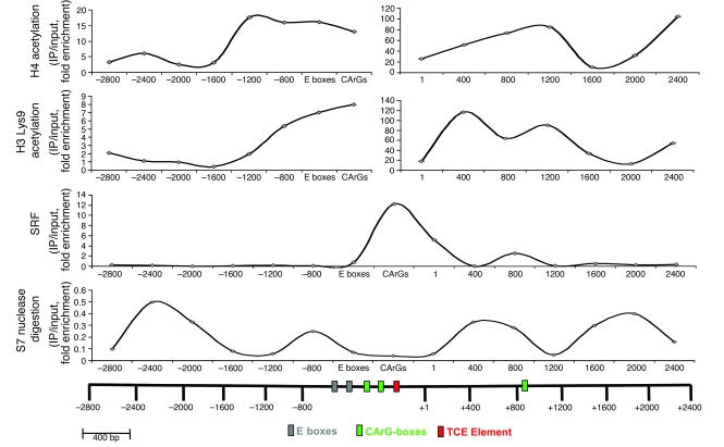
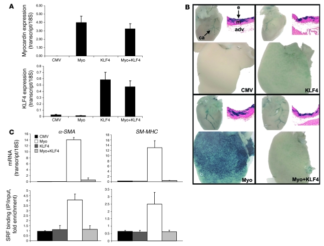
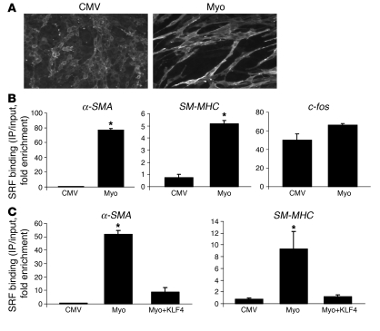
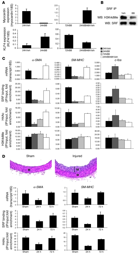
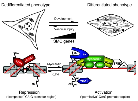
Precise control of SMC transcription plays a major role in vascular development and pathophysiology. Serum response factor (SRF) controls SMC gene transcription via binding to CArG box DNA sequences found within genes that exhibit SMC-restricted expression. However, the mechanisms that regulate SRF association with CArG box DNA within native chromatin of these genes are unknown. Here we report that SMC-restricted binding of SRF to murine SMC gene CArG box chromatin is associated with patterns of posttranslational histone modifications within this chromatin that are specific to the SMC lineage in culture and in vivo, including methylation and acetylation to histone H3 and H4 residues. We found that the promyogenic SRF coactivator myocardin increased SRF association with methylated histones and CArG box chromatin during activation of SMC gene expression. In contrast, the myogenic repressor Kruppel-like factor 4 recruited histone H4 deacetylase activity to SMC genes and blocked SRF association with methylated histones and CArG box chromatin during repression of SMC gene expression. Finally, we observed deacetylation of histone H4 coupled with loss of SRF binding during suppression of SMC differentiation in response to vascular injury. Taken together, these findings provide novel evidence that SMC-selective epigenetic control of SRF binding to chromatin plays a key role in regulation of SMC gene expression in response to pathophysiological stimuli in vivo.


Figures

Figure 1
Figure 1. Cell-specific histone modifications correlate with cell-selective binding of SRF to α-SMA and SM-MHC.
(A) Quantitative ChIP analysis for SRF enrichment at the 5′-CArG boxes of α-SMA, SM-MHC, and c-fos from chromatin isolated from rat aortic SMCs and ECs. (B) Chromatin isolated from SMCs and ECs was digested into mononucleosomal fragments by micrococcal (S7) nuclease. DNA was purified and amplified with primers flanking the 5′-CArG boxes of α-SMA, SM-MHC, and c-fos. The 5′-promoter region of the EC-specific gene VEC was also included as a control. The VEC promoter does not contain CArG boxes. See Methods for explanation. (C) Chromatin was isolated from rat aortas and blood and SRF binding measured by ChIP as in A. (D) Histone modifications that have been associated with activation of gene expression (25) were measured at α-SMA, VEC, cardiac-muscle myosin heavy chain (cardiac-specific; CM α-MHC), and c-fos promoters by ChIP in cultured ES cells (ESC), SMCs, and ECs. We observed patterns similar to α-SMA at SM-MHC (data not shown). All ChIP data for Figure 1 and other figures are plotted as fold enrichment over equivalent amounts of input DNA, and Figure 1D is scaled equally on each horizontal panel (i.e., for each modification) so that enrichments between each of the different promoters can be compared. *P < 0.05 measured by Student’s t test. Controls with beads only and without antibody consistently failed to immunoprecipitate DNA that amplified by real-time PCR (data not shown; see Supplemental Figure 2) for this and all other ChIP experiments. H3K79dMe, H3 Lys79 di-methylation; H3K9Ac, H3 Lys9 acetylation.
Figure 4
Figure 4. KLF4 promotes loss of SRF binding and H4Ac at α-SMA and SM-MHC.
(A) SMCs were infected with CMV-KLF4 (KLF4) or CMV-empty adenoviruses and SRF, H4Ac, and H3K4dMe measured by ChIP. *P < 0.05 by Student’s t test. Accessibility to micrococcal nuclease digestion was measured as in Figure 1B. (B) Cultured SMCs were infected with equivalent amounts of CMV-empty, CMV-myocardin, or CMV-myocardin with CMV-KLF4 (myo+KLF4) adenoviruses and SRF binding to CArG boxes measured by ChIP as above. (C, D, and F) ChIP and real-time RT-PCR were measured from cultured SMCs infected with the corresponding adenoviruses with TSA dissolved in DMSO at 1 ng/ml or 5 ng/ml or in DMSO only. *P < 0.05 for samples when compared with CMV-empty control cells by Student’s t test. (E) SRF was immunoprecipitated from SMCs infected with CMV-KLF4 or control (CMV) viruses, and immunoprecipitates were subjected to Western blotting for H3K4dMe and SRF (IP and IP control).
Figure 3
Figure 3. Identification modifications that contribute to myocardin/SRF binding to CArG boxes.
(A) α-SMA transgenes were stably transfected into rat aortic SMCs by the integrase system as described in Methods, and ChIP was performed with primers specific for the transgene and the endogenous gene. *P < 0.05 by Student’s t test. (B) SMCs were infected with adenovirus harboring CMV-myocardin (Myo) or CMV-empty (CMV) expression vectors, and ChIP was performed for SRF, H4Ac, and H3K4dMe. (C) SMCs were infected with adenovirus as in B. Elk-1, SRF, and FLAG-myocardin immunoprecipitates were subjected to Western blotting for H3K4dMe and SRF. Nonimmune IgG antisera failed to immunoprecipitate SRF and H3K4dMe from SMC extracts in these and all other protein IP experiments (data not shown and Supplemental Figure 2). (D) SMCs were infected as in B, and SRF immunoprecipitates were subjected to Western blotting for H3K4dMe and SRF. (E) SMCs were infected with adenoviruses expressing siRNAs to myocardin (siMyo) or GFP (siGFP; control). Chromatin was isolated, and ChIP measured levels of SRF binding to 5′-CArG boxes. (F) SMCs were infected as in E, and nuclear extracts were treated as in D. (G) Peptide binding assay with FLAG-myocardin as described in Methods. FLAG-myocardin immunoprecipitates collected from SMC extracts containing the corresponding biotinylated peptides were subjected to Western blotting using HRP-streptavidin. H3unmod, unmodified H3 peptide. (H) Myocardin peptide binding assay as in G, comparing the ability of myocardin to immunoprecipitate 2 μg of H3 peptides di-methylated at Lys4 (H3K4dMe), di-methylated at Lys9 (H3K9dMe), acetylated at Lys9, or phosphorylated at serine 10 (H3S10P).
Figure 2
Figure 2. Characterization of transcription factor and histone modification distribution across the α-SMA promoter-enhancer locus.
Fifteen pairs of PCR primers spaced at 400-bp intervals were used in ChIP assays to map the distribution of factors plotted. Nuclease accessibility was determined as in Figure 1. E boxes are cis-elements 5′ to CArG boxes. Data are representative of 2 independent experiments.
Figure 5
Figure 5. Myocardin and KLF4 exert opposing influences over SMC gene expression in transgenic mouse liver in vivo.
(A) mRNA was extracted from liver in mice infected with CMV-empty control viruses, CMV-KLF4, CMV-myocardin, or mice coinjected with myocardin and KLF4 viruses. Expression levels of myocardin and KLF4 were measured by real-time PCR to document that delivery of these genes was successful. Data were normalized to levels of 18S expression. (B) LacZ staining from SM-MHC-LacZ–transgenic mice injected as in A. For each panel in B, the top left image shows hearts expressing SMC-specific LacZ staining in coronary arteries (ca), and the top right image shows cross-sections taken from these mice displaying SMC-specific LacZ staining in the media (m) of aortas. The endothelial layer (e) and adventitia (adv) are labeled. The media exhibit mosaic staining, which is typical for SM-MHC–transgenic mice. The bottom image in each panel shows staining for the presence of LacZ in mouse liver. (C) mRNA levels of α-SMA and SM-MHC were measured by real-time RT-PCR, and SRF binding to CArG box chromatin of these genes was measured by ChIP, in livers of mice injected with the indicated corresponding adenoviruses.
Figure 6
Figure 6. Myocardin and KLF4 exert opposing influences over SRF binding to SMC genes in cultured ECs.
(A) Morphology of ECs infected with CMV-myocardin or control (CMV-empty) adenoviruses. (B) ChIP results for SRF binding to 5′-CArG boxes of the indicated genes from ECs infected with CMV-myocardin adenoviruses or control viruses. (C) ChIP as in B, in ECs infected with CMV-myocardin alone, controls, or ECs coinfected with CMV-myocardin and CMV-KLF4 viruses. *P < 0.05 by student’s t test.
Figure 7
Figure 7. SRF binding to CArG box chromatin is disrupted during SMC phenotypic switching.
(A) Left: Expression of myocardin and KLF4 in response to PDGF-BB (BB) treatment of cultured SMCs for 24 hours (24hBB) or vehicle treatment for 24 hours (24hVeh). Right: Similar data from cells treated with PDGF-BB for 72 hours (72hBB) or cells treated with PDGF-BB media for 24 hours, followed by replacement of PDGF-BB media with vehicle media and incubation for 48 hours (24hBB48hVeh). Data were normalized to expression of 18S. PDGF-BB treatments were performed as described previously (8). (B) SMCs were treated with vehicle (Veh) or PDGF-BB for 24 hours, and SRF immunoprecipitates were subjected to Western blotting for H3K4dMe or SRF (IP control). (C) SMCs were treated with PDGF-BB as in A, mRNA was measured by RT-PCR as in A (top panel), and ChIP was performed at the 5′-CArG regions with the indicated genes for the indicated parameters (SRF binding, etc). (D) Rats were injured with balloon catheter as described in Methods, and mRNA or chromatin was isolated and analyzed by real-time RT-PCR and ChIP, respectively. The top panel is a histological display of uninjured control aorta (Sham) and an injured aorta (Injured), demonstrating that the balloon catheter injury technique employed successfully injured the vessels, as indicated by the presence of a neointima (NI) 14 days after injury. M refers to the position of the vessel media. For mRNA and ChIP (bottom panels), vessels were harvested 24 hours or 72 hours after injury and compared with control vessels.
Figure 8
Figure 8. Model for epigenetic regulation of SRF binding to CArG box chromatin.
Gray squares represent histone octamers with red DNA strands wrapped around them. The dark lines with Me (methyl groups) and Ac (acetyl groups) protruding from H3 and H4 represent histone tail domains that are subject to H4 and H3 acetylation and H3 Lys4 methylation. In this model, signals such as vascular injury that repress myocardin and/or recruit KLF4-dependent HDAC activity at SMC gene promoters result in loss of SRF binding and transcriptional repression of these genes, to promote the dedifferentiated phenotype. In contrast, in the absence of KLF4, SRF is able to recognize accessible CArG box sequences within “open” chromatin containing H4Ac, synergizing with docking of myocardin to H3K4dMe, to facilitate SRF binding to chromatin and transcriptional activation, which promotes SMC differentiation. The blue protein labeled “??” represents a putative myocardin-accessory factor that may assist myocardin in docking to methylated histones near CArG DNA sequences, to help tether and/or stabilize SRF binding to SMC gene chromatin, which is enriched with H3K4dMe in SMCs.

References

    1. Owens G.K., Kumar M.S., Wamhoff B.R. Molecular regulation of vascular smooth muscle cell differentiation in development and disease. Physiol. Rev. 2004;84:767–801. - PubMed
    1. Hoofnagle M.H., Wamhoff B.R., Owens G.K. Lost in transdifferentiation. J. Clin. Invest. 2004;113:1249–1251. - PMC - PubMed
    1. Hu Y., et al. Abundant progenitor cells in the adventitia contribute to atherosclerosis of vein grafts in ApoE-deficient mice. J. Clin. Invest. 2004;113:1258–1265. - PMC - PubMed
    1. Sata M., et al. Hematopoietic stem cells differentiate into vascular cells that participate in the pathogenesis of atherosclerosis. Nat. Med. 2002;8:403–409. - PubMed
    1. Glaser R., Lu M.M., Narula N., Epstein J.A. Smooth muscle cells, but not myocytes, of host origin in transplanted human hearts. Circulation. 2002;106:17–19. - PubMed