. 2005 Dec 7;33(21):6795-804.
doi: 10.1093/nar/gki982. Print 2005.

MPP6 is an exosome-associated RNA-binding protein involved in 5.8S rRNA maturation

Affiliations

MPP6 is an exosome-associated RNA-binding protein involved in 5.8S rRNA maturation

Geurt Schilders et al. Nucleic Acids Res. .

Abstract

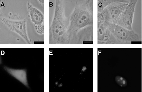
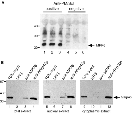
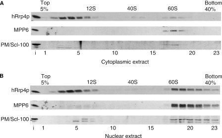
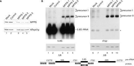
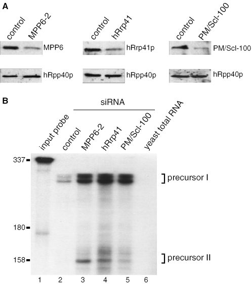
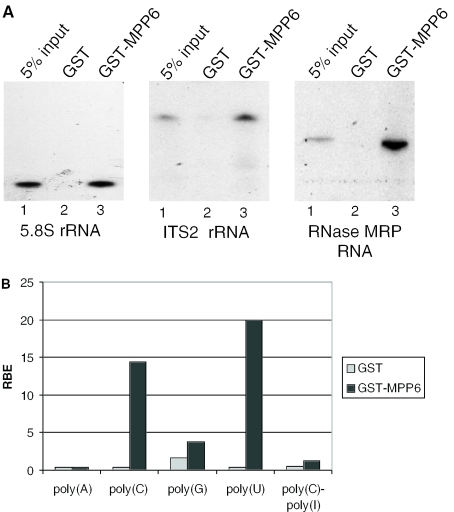
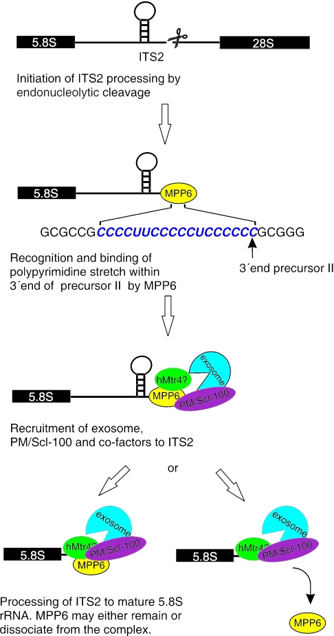
The exosome is a complex of 3'-->5' exoribonucleases which is involved in many RNA metabolic processes. To regulate these functions distinct proteins are believed to recruit the exosome to specific substrate RNAs. Here, we demonstrate that M-phase phosphoprotein 6 (MPP6), a protein reported previously to co-purify with the TAP-tagged human exosome, accumulates in the nucleoli of HEp-2 cells and associates with a subset of nuclear exosomes as evidenced by co-immunoprecipitation and biochemical fractionation experiments. In agreement with its nucleolar accumulation, siRNA-mediated knock-down experiments revealed that MPP6 is involved in the generation of the 3' end of the 5.8S rRNA. The accumulation of the same processing intermediates after reducing the levels of either MPP6 or exosome components strongly suggests that MPP6 is required for the recruitment of the exosome to the pre-rRNA. Interestingly, MPP6 appeared to display RNA-binding activity in vitro with a preference for pyrimidine-rich sequences, and to bind to the ITS2 element of pre-rRNAs. Our data indicate that MPP6 is a nucleolus-specific exosome co-factor required for its role in the maturation of 5.8S rRNA.


Figures

Figure 1
Figure 1
Subcellular localization of MPP6. HEp-2 cells were transfected with constructs encoding EGFP alone (A and D), EGFP-MPP6 (B and E) and EGFP-hMtr3p (C and F) and after 24 h the cells were fixed in 4% paraformaldehyde/PBS and the expressed fusion proteins were analysed by fluorescence microscopy. Phase-contrast and fluorescence images are displayed in (A–C and D–F), respectively. Each bar corresponds to 10 µm.
Figure 2
Figure 2
MPP6 is associated with nuclear exosome complexes. (A) HEp-2 cell extracts were subjected to immunoprecipitation with three anti-PM/Scl-positive (lanes 1–3) and three anti-PM/Scl-negative patient sera (lanes 4–6). Precipitated proteins were analysed by western blotting using a rabbit anti-MPP6 serum. The positions of molecular mass markers (kDa) are indicated on the left. (B) Reciprocal experiment in which a normal rabbit serum (lanes 2, 6 and 10), a polyclonal anti-MPP6 serum (lanes 3, 7 and 11) and a polyclonal anti-hRrp40p serum (lanes 4, 8 and 12) were used for immunoprecipitations. A monoclonal antibody to hRrp4p was used for the detection of a co-precipitated exosome component. For the immunoprecipitations total (lanes 1–4), nuclear (lanes 5–8) and cytoplasmic (lanes 9–12) HEp-2 cell extracts were used. Input material (10%) of the extracts was loaded in lanes 1, 5 and 9, respectively. The positions of molecular mass markers (kDa) are indicated on the left.
Figure 3
Figure 3
MPP6 predominantly co-sediments with nuclear exosome complexes at 60S–80S in glycerol gradients. Cytoplasmic (A) and nuclear (B) extracts from HEp-2 cells were fractionated in a 5–40% (v/v) glycerol gradient. The sedimentation of hRrp4, MPP6 and PM/Scl-100 was determined by immunoblotting. The sedimentation of the large rRNAs was determined by agarose gel electrophoresis and ethidium bromide staining and used as markers (40S and 60S) in the gradient. U1 snRNA was used as marker for 12S complexes.
Figure 4
Figure 4
Knock down of MPP6 by RNAi leads to the accumulation of 5.8S rRNA precursors. (A) HEp-2 cells were transiently transfected with three different siRNAs to MPP6 (100 pmol), control siRNA or buffer (mock). Cells were harvested 2 days after transfection and 75 µg of total protein was analysed by western blotting using a polyclonal anti-MPP6 serum or a polyclonal anti-hRrp41p serum (control). (B) Northern analysis of 5.8S rRNA processing upon MPP6 knock down. Total RNA (5 µg) from (mock) transfected cells was analysed by northern blot hybridization using radiolabelled probes specific for 5.8S rRNA (left) or ITS2 (right). The relative positions of these probes, and also the other probes used, with respect to the primary rRNA transcript are depicted in the scheme below the autoradiographs. Note that the size of the probes is not proportional to that of the pre-rRNA. As a control, a U6 snRNA probe was used. The positions of 5.8S rRNA and its precursors (I and II) are indicated. The asterisk in the right panel points to a weak cross-hybridization of the ITS2 probe with the mature 5.8S rRNA.
Figure 5
Figure 5
MPP6, hRrp41 and PM/Scl-100 knock down lead to the accumulation of the same 5.8S rRNA precursors. (A) HEp-2 cells were transiently transfected with control siRNA (100 pmol), MPP6-2 siRNA (100 pmol), hRrp41 siRNA (200 pmol) and PM/Scl-100 siRNA (100 pmol). After 36 h transfection cells were retransfected with the same amount of siRNA and after an additional 36 h the cells were harvested. The proteins were analysed by western blotting using 75 µg of total protein extracts from transfected cells and a polyclonal anti-MPP6 serum, a polyclonal anti-hRrp41 serum, and a polyclonal anti-PM/Scl-100 serum. A polyclonal anti-hRpp40 serum was used as a control. (B) A radiolabelled antisense ITS2 probe, complementary to the first 300 nt of ITS2, was hybridized to 5 µg of total RNA from siRNA-treated cells. After digestion with RNase A and RNase T1 the RNA was analysed on a 10% denaturing polyacrylamide gel. The radiolabelled ITS2 probe was loaded in lane 1. As a control, the same probe was incubated with 5 µg of yeast total RNA and further treated by the same procedure (lane 6). Lanes 4–6 show protected RNAs from cells treated with control, MPP6-2, Rrp41 and PM/Scl-100 siRNA, respectively. The positions of the 5.8S precursor rRNAs (I and II) are indicated. Positions of marker RNAs are indicated on the left.
Figure 6
Figure 6
GST-tagged MPP6 binds to in vitro transcribed RNAs and prefers polypyrimidines. (A) GST and GST-MPP6 recombinant proteins were incubated with radiolabeled, in vitro transcribed full-length 5.8S rRNA, ITS2 rRNA (a fragment corresponding to the most 5′ 300 nt) and RNase MRP RNA. Binding to these RNAs was assayed by GST pull-down followed by denaturing gel electrophoresis and autoradiography. Five percent of the input RNA was loaded in lanes 1. Lanes 2 and 3 contain the material precipitated by GST alone and GST-MPP6, respectively. (B) Binding of GST and GST–MPP6 fusion proteins to radiolabelled homopolynucleotides was analysed as described above and the bound RNAs were quantified in a scintillation counter. The binding efficiency of GST or GST-MPP6 with poly(A), poly(C), poly(G), poly(U) and poly(C)–ply(I) is depicted as a percentage of input RNA (RBE: relative binding efficiency). These results are the averages of two independent experiments.
Figure 7
Figure 7
Model for the role of MPP6 in pre-rRNA processing. Before, or directly after cleavage of the pre-rRNA in ITS2 by a yet unknown endoribonuclease, MPP6 binds to oligopyrimidine stretches in the ITS2 RNA and subsequently recruits the PM/Scl-100-containing exosome and probably several additional factors, like hMtr4p or an hMtr4p containing complex. This is in agreement with the reported two-hybrid interactions between MPP6 and PM/Scl-100 and between MPP6 and hMtr4p (25). The exosome will then process the ITS2 RNA to generate the mature 5.8S rRNA. MPP6 may either remain associated with the processing complex or dissociate once the exosome starts the digestion.

Similar articles

Cited by

References

    1. Mitchell P., Petfalski E., Shevchenko A., Mann M., Tollervey D. The exosome: a conserved eukaryotic RNA processing complex containing multiple 3′→5′ exoribonucleases. Cell. 1997;91:457–466. - PubMed
    1. Mitchell P., Petfalski E., Tollervey D. The 3′ end of yeast 5.8S rRNA is generated by an exonuclease processing mechanism. Genes Dev. 1996;10:502–513. - PubMed
    1. Allmang C., Kufel J., Chanfreau G., Mitchell P., Petfalski E., Tollervey D. Functions of the exosome in rRNA, snoRNA and snRNA synthesis. EMBO J. 1999;18:5399–5410. - PMC - PubMed
    1. Kufel J., Allmang C., Chanfreau G., Petfalski E., Lafontaine D., Tollervey D. Precursors to the U3 small nucleolar RNA lack small nucleolar RNP proteins but are stabilized by La binding. Mol. Cell. Biol. 2000;20:5415–5424. - PMC - PubMed
    1. Mitchell P., Petfalski E., Houalla R., Podtelejnikov A., Mann M., Tollervey D. Rrp47p is an exosome-associated protein required for the 3′ processing of stable RNAs. Mol. Cell. Biol. 2003;23:6982–6992. - PMC - PubMed

Publication types

MeSH terms