

Down-regulation of interferon regulatory factor 4 gene expression in leukemic cells due to hypermethylation of CpG motifs in the promoter region
- PMID: 16396836
- PMCID: PMC1310901
- DOI: 10.1093/nar/gki1001
Down-regulation of interferon regulatory factor 4 gene expression in leukemic cells due to hypermethylation of CpG motifs in the promoter region
Abstract
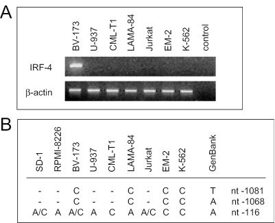
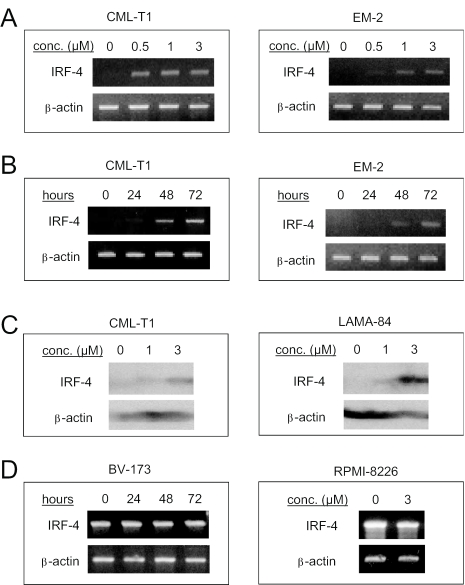
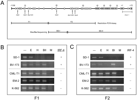
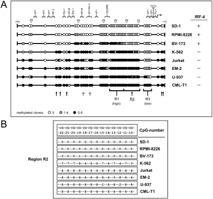
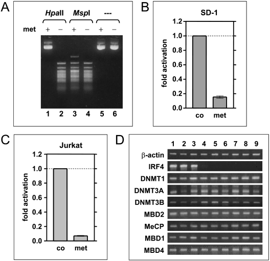
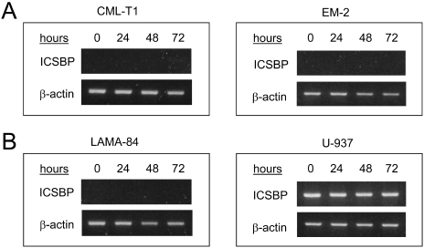
Although the bcr-abl translocation has been shown to be the causative genetic aberration in chronic myeloid leukemia (CML), there is mounting evidence that the deregulation of other genes, such as the transcription factor interferon regulatory factor 4 (IRF-4), is also implicated in the pathogenesis of CML. Promoter methylation of CpG target sites or direct deletions/insertions of genes are mechanisms of a reversible or permanent silencing of gene expression, respectively. Therefore, we investigated whether IRF-4 promoter methylation or mutation may be involved in the regulation of IRF-4 expression in leukemia cells. Whereas promoter mutations or structural rearrangements could be excluded as a cause of altered IRF-4 expression in hematopoietic cells, the IRF-4 promoter methylation status was found to significantly influence IRF-4 transcription. First, treatment of IRF-4-negative lymphoid, myeloid and monocytic cell lines with the methylation-inhibitor 5-aza-2-deoxycytidine resulted in a time- and concentration-dependent increase of IRF-4 mRNA and protein levels. Second, using a restriction-PCR-assay and bisulfite-sequencing we identified specifically methylated CpG sites in IRF-4-negative but not in IRF-4-positive cells. Third, we clearly determined promoter methylation as a mechanism for IRF-4 down-regulation via reporter gene assays, but did not detect an association of methylational status and mRNA expression of DNA methyltransferases or methyl-CpG-binding proteins. Together, these data suggest CpG site-specific IRF-4 promoter methylation as a putative mechanism of down-regulated IRF-4 expression in leukemia.
Figures
Similar articles
-
Correlation between the single-site CpG methylation and expression silencing of the XAF1 gene in human gastric and colon cancers.Gastroenterology. 2006 Dec;131(6):1835-43. doi: 10.1053/j.gastro.2006.09.050. Epub 2006 Oct 1. Gastroenterology. 2006. PMID: 17087954
-
[Down-regulation of transcription factor PU.1 via abnormal epigenetic modification in chronic myeloid leukemia].Zhonghua Zhong Liu Za Zhi. 2012 Mar;34(3):169-75. doi: 10.3760/cma.j.issn.0253-3766.2012.03.003. Zhonghua Zhong Liu Za Zhi. 2012. PMID: 22780968 Chinese.
-
Methylation silencing of the Apaf-1 gene in acute leukemia.Mol Cancer Res. 2005 Jun;3(6):325-34. doi: 10.1158/1541-7786.MCR-04-0105. Mol Cancer Res. 2005. PMID: 15972851
-
Regulation of CpG methylation by Dnmt and Tet in pluripotent stem cells.J Reprod Dev. 2016 Aug 25;62(4):331-5. doi: 10.1262/jrd.2016-046. Epub 2016 May 5. J Reprod Dev. 2016. PMID: 27151232 Free PMC article. Review.
-
miRNA and methylation: a multifaceted liaison.Chembiochem. 2015 Jan 19;16(2):195-203. doi: 10.1002/cbic.201402449. Epub 2014 Dec 2. Chembiochem. 2015. PMID: 25469751 Review.
Cited by
-
IRF-4 functions as a tumor suppressor in early B-cell development.Blood. 2008 Nov 1;112(9):3798-806. doi: 10.1182/blood-2007-10-117838. Epub 2008 Aug 19. Blood. 2008. PMID: 18713947 Free PMC article.
-
Evaluation of a panel of tumor-specific differentially-methylated DNA regions in IRF4, IKZF1 and BCAT1 for blood-based detection of colorectal cancer.Clin Epigenetics. 2021 Jan 21;13(1):14. doi: 10.1186/s13148-020-00999-y. Clin Epigenetics. 2021. PMID: 33478584 Free PMC article.
-
Promoter methylation and expression levels of selected hematopoietic genes in pediatric B-cell acute lymphoblastic leukemia.Blood Res. 2015 Mar;50(1):26-32. doi: 10.5045/br.2015.50.1.26. Epub 2015 Mar 24. Blood Res. 2015. PMID: 25830127 Free PMC article.
-
The IRF family of transcription factors: Inception, impact and implications in oncogenesis.Oncoimmunology. 2012 Nov 1;1(8):1376-1386. doi: 10.4161/onci.22475. Oncoimmunology. 2012. PMID: 23243601 Free PMC article.
-
The dynamic functions of IRF4 in B cell malignancies.Clin Exp Med. 2023 Aug;23(4):1171-1180. doi: 10.1007/s10238-022-00968-0. Epub 2022 Dec 10. Clin Exp Med. 2023. PMID: 36495369 Free PMC article. Review.
References
-
- Kantarjian H., O'Brien S., Anderlini P., Talpaz M. Treatment of myelogenous leukemia: current status and investigational options. Blood. 1996;87:3069–3081. - PubMed
-
- Sawyers C.L. Chronic myeloid leukemia. N. Engl. J. Med. 1999;340:1330–1340. - PubMed
-
- Schmidt M., Hochhaus A., König-Merediz S.A., Brendel C., Proba J., Hoppe G.J., Wittig B., Ehninger G., Hehlmann R., Neubauer A. Expression of interferon regulatory factor 4 in chronic myeloid leukemia: correlation with response to interferon-α therapy. J. Clin. Oncol. 2000;18:3331–3338. - PubMed
-
- Nguyen H., Hiscott J., Pitha P.M. The growing family of interferon regulatory factors. Cytokine Growth Factor Rev. 1997;8:293–312. - PubMed
-
- Tanaka N., Taniguchi T. The interferon regulatory factors and oncogenesis. Semin. Cancer Biol. 2000;10:73–81. - PubMed
Publication types
MeSH terms
Substances
LinkOut - more resources
Full Text Sources
Other Literature Sources
Medical
Miscellaneous
