. 2006 Apr 15;107(8):3350-8.
doi: 10.1182/blood-2005-09-3556. Epub 2006 Jan 5.

Mutation of mouse Mayp/Pstpip2 causes a macrophage autoinflammatory disease

Affiliations

Mutation of mouse Mayp/Pstpip2 causes a macrophage autoinflammatory disease

Johannes Grosse et al. Blood. .

Abstract

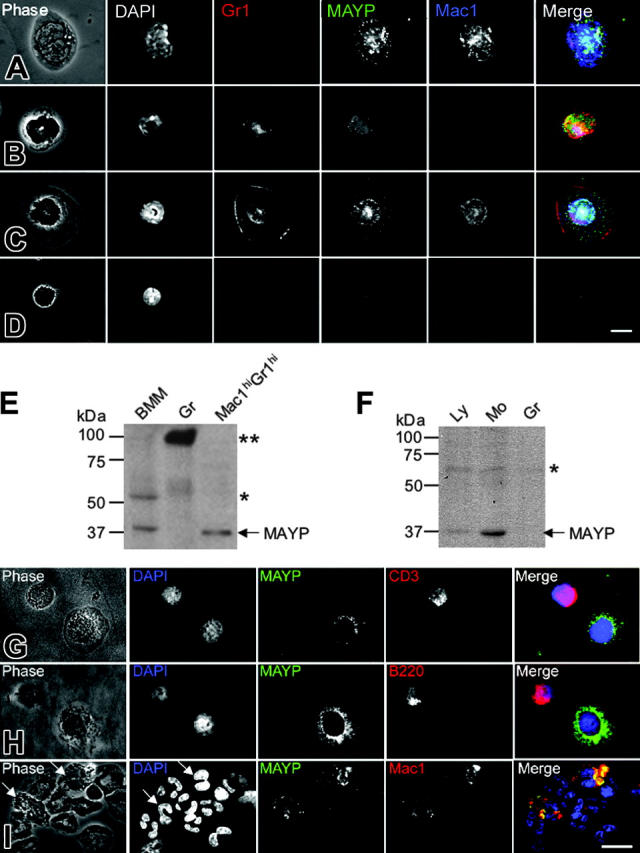
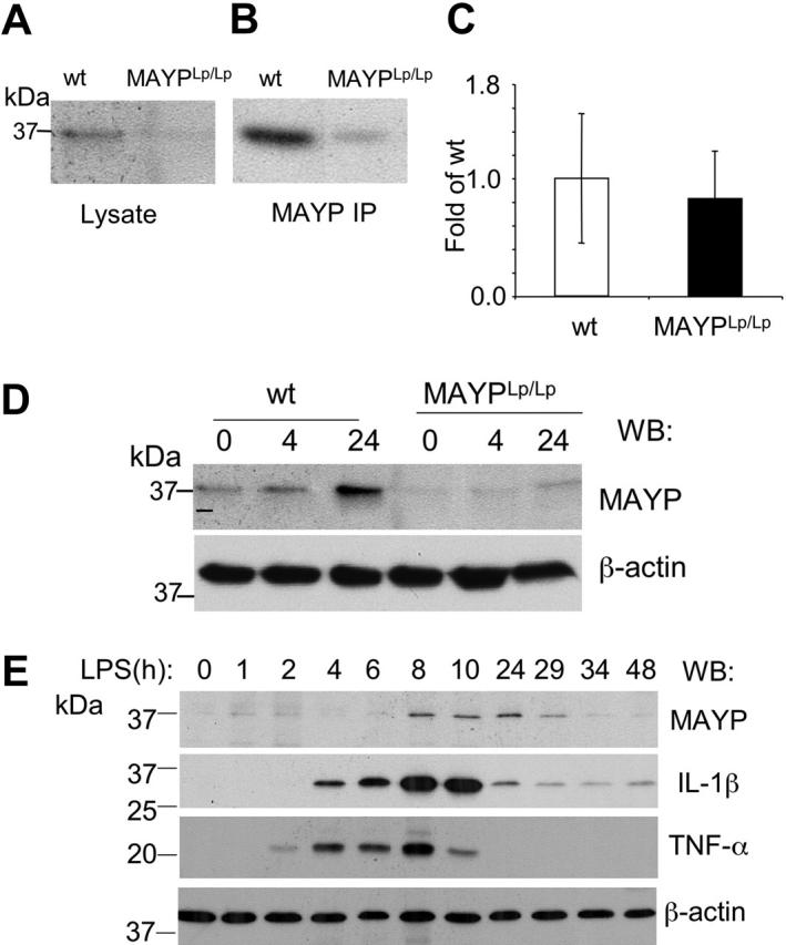
Macrophage actin-associated tyrosine phosphorylated protein (MAYP)/PSTPIP2, a PCH protein, is involved in the regulation of macrophage motility. Mutations in a closely related gene, PSTPIP1/CD2BP1, cause a dominantly inherited autoinflammatory disorder known as PAPA syndrome. A mutant mouse obtained by chemical mutagenesis exhibited an autoinflammatory disorder characterized by macrophage infiltration and inflammation, leading to osteolysis and necrosis in paws and necrosis of ears. Positional cloning of this recessive mutation, termed Lupo, identified a T to A nucleotide exchange leading to an amino acid substitution (I282N) in the sequence of MAYP. Mayp(Lp/Lp) disease was transferable by bone marrow transplantation and developed in the absence of lymphocytes. Consistent with the involvement of macrophages, lesion development could be prevented by the administration of clodronate liposomes. MAYP is expressed in monocytes/macrophages and in a Mac1+ subfraction of granulocytes. LPS stimulation increases its expression in macrophages. Because of the instability of the mutant protein, MAYP expression is reduced 3-fold in Mayp(Lp/Lp) macrophages and, on LPS stimulation, does not rise above the level of unstimulated wild-type (WT) cells. Mayp(Lp/Lp) mice expressed elevated circulating levels of several cytokines, including MCP-1; their macrophages exhibited altered cytokine production in vitro. These studies suggest that MAYP plays an anti-inflammatory role in macrophages.


Figures

Figure 1.
Figure 1.
Clinical symptoms and disease progression in homozygous Lupo mice. (A-B) Early clinical symptoms with edematous inflammation, proceeding to ulceration, crust formation, and necrosis of 2 digits (B, arrows). (C) Ear destruction by ulcerative inflammation. (D) Radiogram showing the destruction of the distal phalanx of digit 3 in a homozygous mutant mouse. Note the shadow caused by the surrounding edematous soft tissue (arrow). (E) Radiogram of WT control paw. (F) Cumulative fraction free of clinical events for Lupo homozygotes on the C3H background, in outcross/intercross (IC) Rag1+/+, C3H/BL6 (50%/50%) population used for positional cloning and on the Rag1-/-, C3H/BL6 (50%/50%) background. On the Rag1+/+, C3H/BL6 background, the median age of onset was significantly later, and lifetime penetrance was lower than on the C3H or the Rag1-/-, C3H/BL6 background (Kaplan-Meier with log rank test [P < .001] for C3H/BL6 versus C3H and C3H/BL6 versus Rag1-/-, C3H/BL6; censored, termination of observation without an event).
Figure 2.
Figure 2.
Histology of dermis and paws of homozygous Lupo mice. (A-B) Hematoxylin and eosin staining of an early stage of dermal inflammation. Inflammatory infiltration is restricted to the connective tissue of the corium and the epithelium (arrowheads, A) and paws (B), showing inflammation starting from the corium (arrow) and spreading to erode the articular cartilage (arrowheads). (C-D) Low-power (C) and high-power (D) images of ears of Lupo (left panels) and WT (right panels) mice, immunostained for the macrophage marker, F4/80+, and counterstained with hematoxylin, showing increased ear thickness, hyperkeratosis, acanthotic epidermis, and increased macrophage infiltration in the external ears of the Lupo mice. (E-F) Low-power (E) and high-power (F) images of paws of Lupo (left panels) and WT (right panels) mice, immunostained for the macrophage marker F4/80+ and counterstained with hematoxylin, showing increased paw thickness, acanthotic epidermis, and increased macrophage infiltration in the footpads of the Lupo mice. Bars, 100 μm.
Figure 3.
Figure 3.
Mapping of the Lupo mutation. (A) Genomic structure of the mapping region on chromosome 18. Locations of flanking and cosegregating markers are indicated above the chromosomal ruler and the genes below it. Positions of genes Ccdc5, Atp5a1, Slc14a1, Slc14a2, Setbp1, and Pstpip2/Mayp and the mapped EST clones 8030462N17Rik and AK028969/AK122536 are shown. (B) Haplotypes of the informative mice (IC1 and IC2) define the mapping region.
Figure 4.
Figure 4.
Elevation of circulating immunoglobulin and autoantibodies in 4-month-old MaypLp/Lp mice. (A) Serum immunoglobulin levels expressed as the fold increase of their concentration in MaypLp/Lp (n = 5) compared with WT (n = 17) mice (± SD). Elevations of IgG1, IgG2a, and IgE are significant (P < .05; Student t test). (B) Twelve lanes, each containing identical amounts of a single extract of normal paw proteins, were subjected to SDS-PAGE and Western blot analysis with individual sera of 6 MaypLp/Lp mice expressing serum immunoglobulin and of 6 WT mice. (C) Immunohistochemistry using serum from a MaypLp/Lp mouse as primary antibody, showing that the cognate proteins are located in the papillary layers of the corium. Similar results were obtained for 3 additional MaypLp/Lp sera. Bar, 100 μm.
Figure 5.
Figure 5.
MAYP is expressed in macrophages and a subset of granulocytes in mice and humans. (A-D) Immunofluorescence staining of mouse peripheral blood leukocytes FACS-sorted based on Mac1 and Gr1 and cytocentrifuged onto fibronectin-coated coverslips, fixed, permeabilized, and stained with DAPI and anti-MAYP antibody, showing the pattern of MAYP staining in Mac1hiGr1lo monocytes (A), Mac1loGr1hi granulocytes (B), Mac1hiGr1hi monocytes or monocyte precursors (C), and Mac1loGr1lo lymphocytes (D). Note diffuse staining of MAYP in panel B and its exclusively nuclear staining in panel D compared with the punctate cytoplasmic staining characteristic of MAYP in panels A and C. Panels on the right represent merged signals of Mac-1 (blue), MAYP (green), and Gr1 (red). (E) SDS-PAGE and Western blot analysis of MAYP expression in mouse BMMs, granulocytes (Gr), and the Mac1hiGr1hi fraction. (F) Western blot analysis of MAYP expression in lymphocyte (Ly; nonadherent peripheral blood mononuclear cells), monocyte (Mo; adherent peripheral blood mononuclear cells), and granulocyte (Gr) fractions of human peripheral blood. (E-F) Single and double asterisks indicate positions of cross-reactive protein bands. (G-I) Immunofluorescence staining of fractions from panel F. Ly fraction (G-H), showing contaminating MAYP+ monocytes, and Gr fraction (I) showing MAYP staining of a Mac1+ subfraction of the granulocytes (10% of total). Bars, 10 μm.
Figure 6.
Figure 6.
Reduced MAYP expression in MaypLp/Lp macrophages. SDS-PAGE and Western blot analysis of MAYP expression in NP-40 cell lysates (A) and anti-MAYP immunoprecipitates (B) prepared from BMMs of MaypLp/Lp and WT mice. (C) Similar levels of expression of MAYP mRNA in BMMs of MaypLp/Lp and WT mice determined by quantitative RT-PCR (± SD, triplicate assays, 2 mice per genotype). (D) LPS stimulation for 4 or 24 hours fails to elevate MAYP expression in MaypLp/Lp BMMs above the level of its expression in unstimulated WT BMMs. (E) Peak response of MAYP expression to LPS follows peak responses of the proinflammatory cytokines IL-1β and TNF-α. β-Actin is the loading control.
Figure 7.
Figure 7.
Differences in cytokine production by WT and MaypLp/Lp macrophages. (A) Cytokine release by cultured WT (formula image
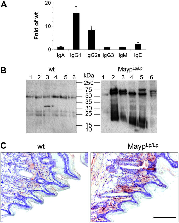
) and MaypLp/Lp (□) BMM. (B) Cytokine release in WT (formula image) and MaypLp/Lp (□) BMMs stimulated with LPS for 24 hours. Duplicate measurements for BMMs from 2 mice of each genotype. Differences between WT and MaypLp/Lp are significant (P < .05; Student t test; n = 4). No significant differences were seen for the other 34 cytokines tested and listed in “Materials and methods.”

Similar articles

Cited by

References

    1. Galon J, Aksentijevich I, McDermott MF, O'Shea JJ, Kastner DL. TNFRSF1A mutations and autoinflammatory syndromes. Curr Opin Immunol. 2000;12: 479-486. - PubMed
    1. McDermott MF, Aksentijevich I, Galon J, et al. Germline mutations in the extracellular domains of the 55 kDa TNF receptor, TNFR1, define a family of dominantly inherited autoinflammatory syndromes. Cell. 1999;97: 133-144. - PubMed
    1. Hull KM, Drewe E, Aksentijevich I, et al. The TNF receptor-associated periodic syndrome (TRAPS): emerging concepts of an autoinflammatory disorder. Medicine (Baltimore). 2002;81: 349-368. - PubMed
    1. McDermott MF. A common pathway in periodic fever syndromes. Trends Immunol. 2004;25: 457-460. - PubMed
    1. Spencer S, Dowbenko D, Cheng J, et al. PSTPIP: a tyrosine phosphorylated cleavage furrow-associated protein that is a substrate for a PEST tyrosine phosphatase. J Cell Biol. 1997;138: 845-860. - PMC - PubMed

Publication types

MeSH terms