. 2006 Jan 1;66(1):259-66.
doi: 10.1158/0008-5472.CAN-05-2502.

Matrix metalloproteinase-9 from bone marrow-derived cells contributes to survival but not growth of tumor cells in the lung microenvironment

Affiliations

Matrix metalloproteinase-9 from bone marrow-derived cells contributes to survival but not growth of tumor cells in the lung microenvironment

Heath B Acuff et al. Cancer Res. .

Abstract

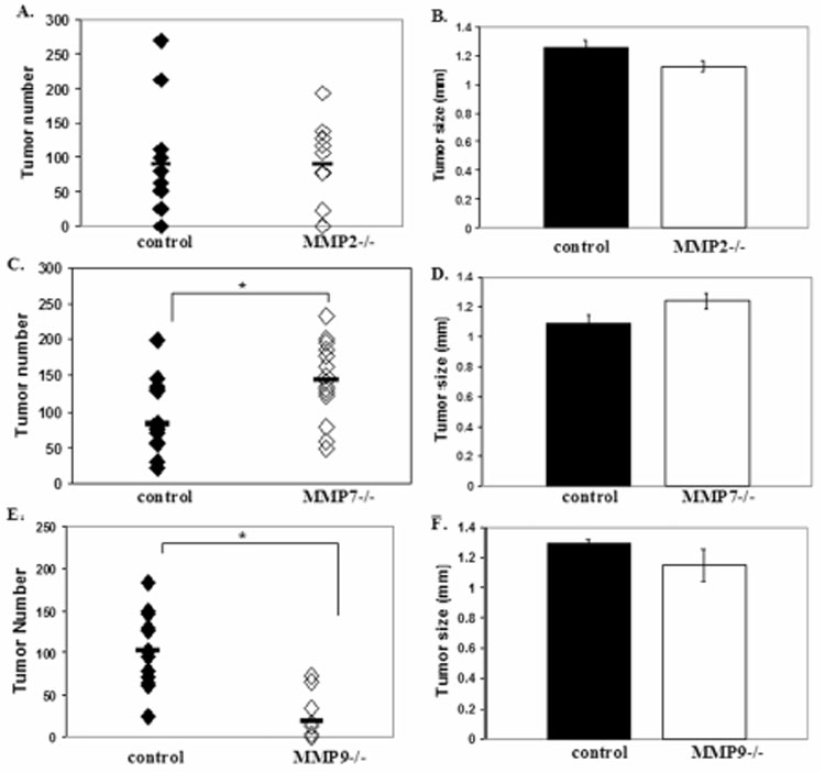
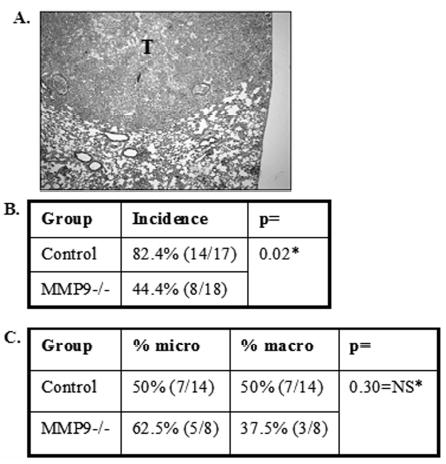
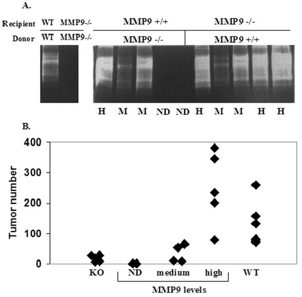
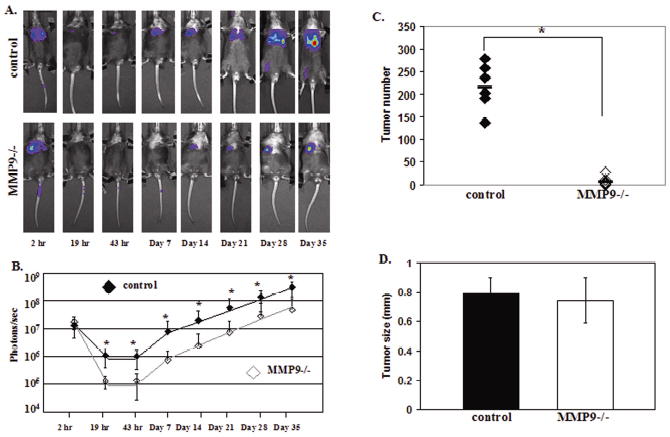
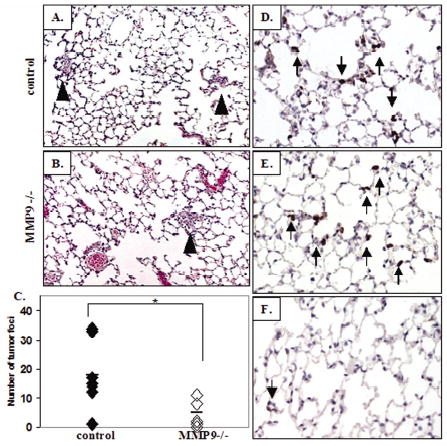
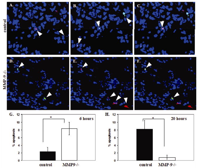
The role of specific stromal-derived matrix metalloproteinases (MMPs) was analyzed in experimental metastasis assays in wild-type and either MMP-9, MMP-7, or MMP-2 null mice. MMP-9 null mice showed an 81% reduction in Lewis lung carcinoma tumor number, whereas MMP-7 null mice showed a 42% increase in tumor number, and there was no difference in tumor number in MMP-2 null mice compared with wild-type controls. Similarly, in an orthotopic model of lung cancer, 50% fewer MMP-9 null mice were able to establish tumors in the lung compared with control mice, although the size of the tumors was not different. The effect of MMP-9 on lung tumor colonization was dependent on the expression of MMP-9 from bone marrow-derived cells and is most likely contributed by neutrophils. To examine temporal effects of stromal MMP-9, bioluminescence imaging from luciferase-expressing human lung cancer-derived A549 cells revealed that there were fewer tumor cells in the lungs of MMP-9 null mice as early as 19 hours after injection compared with control mice, with no difference in subsequent growth rates. Six hours after injection of tumor cells, MMP-9 null mice showed a 4-fold increase in the percent of tumor cells undergoing apoptosis compared with control mice. We conclude that MMP-9 from the bone marrow contributes to the early survival and establishment of tumors in the lung and has no effect on subsequent growth. These results provide insights into the failure of MMP inhibitors in clinical trials in patients with late-stage lung cancer.


Figures

Figure 1
Figure 1
Host MMP contributions to lung tumor formation. (A) Number of surface lung tumors in control (n=12) and MMP2 null (n=12) mice 2 weeks after injection of 3 x 10⁵ LLC cells i.v. (p=NS). (B) Tumor size in control and MMP2 null mice (p=NS). (C) Number of surface lung tumors in control (n=14) and MMP7 null (n=14) mice 2 weeks after injection of 3 x 10⁵ LLC cells i.v. (*p=0.01). (D) Tumor size in control and MMP7 null mice (p=NS). (E) Number of surface lung tumors in control (n=13) and MMP9 null (n=11) mice 2 weeks after injection of 3 x 10⁵ LLC cells i.v. (*p=0.0002). (F) Tumor size in control and MMP9 null mice (p=NS).
Figure 2
Figure 2
Host MMP9 contributes to establishment of a primary tumor in the lung. (A) Histology of lung 5 weeks after orthotopic injection of 1 x 10⁶ A549 cells (H/E). T= tumor. Table B: Percent of control (n=17) and MMP9 null mice (n=18) that establish a primary tumor in the lung after orthotopic injection of A549 cells (*p=0.02). Table C: Percentage of tumors in control and MMP9−/− mice that were classified as microscopic (micro) (<0.1 mm3) or macroscopic (macro) (0.5–87.5 mm3). *Fisher’s exact test-2-tailed.
Figure 3
Figure 3
MMP9 levels from bone marrow-derived cells correlate with lung tumor formation. (A) Gelatin zymography of bone marrow cell lysates showing MMP levels in representative samples of wildtype (WT) and MMP9 null (MMP−/−) mice after reconstitution with either MMP9 positive or MMP9 null bone marrow cells. ND=not detected, M=medium, H=high levels of bone marrow MMP9. Duplicate zymograms in the presence of EDTA confirms metal-dependent enzymatic activity. (B) Quantitation of surface lung tumors in control or MMP9 null mice reconstituted with MMP9 positive or MMP9 null bone marrow cells.
Figure 4
Figure 4
MMP9 contributes to the early establishment of tumors in the lung. (A) In vivo bioluminescent images of representative control and MMP9 null mice injected with 2x10⁶ luciferase expressing A549 cells (LUC-A549) i.v. into the tail vein. (B) Mean ± SD tumor growth rates in control (n=10) and MMP9 null (n=10) mice at the time points indicated in (A) determined by bioluminescence (photons/sec) from the LUC-A549 cells (*p <0.05). (C) Number of surface lung tumors in control (n=6) and MMP9 null (n=10) mice 5 weeks after injection of 2 x 10⁶ LUC-A549 cells i.v. (* p=0.0007) (D) Surface lung tumor size (mm) in control and MMP9 null mice (p=NS).
Figure 5
Figure 5
MMP9 null mice show fewer tumor cells in the lung 24 hours after tumor cell inoculation compared to control mice and both groups show neutrophil infiltration. Histological (H/E) analysis of lungs in control (A) and MMP9 null (B) mice 24 hours after i.v. injection of A549 cells into the tail vein. Clusters of tumor cells are indicated by arrow heads. (C) Quantitation of tumor cell clusters in the lungs of control (n=7) and MMP9 null mice (n=6) 24 hours after tumor cell inoculation (*p=0.018). Immunohistochemical analysis of neutrophils 24 hours after tumor cell inoculation in control (D) and MMP9 null (E) mice. Arrows indicate neutrophils in the lungs. (F) Neutrophils in the lungs of non-injected Rag2 null mice.
Figure 6
Figure 6
MMP9 null mice show significantly more tumor cells undergoing apoptosis 6 hours after injection of tumor cells. (A–G) TUNEL analysis at 6 hours after injection of CellTracker labeled A549 cells in control (A–C) and MMP9 null (D–F) mice. Apoptotic cells (green, A and D). CellTracker labeled tumor cells (red, B and E). C and F are the localization of apoptotic tumor cells shown in yellow by a merge of TUNEL staining (green) and CellTracker staining (red). Nuclei are blue (Hoechst). (G) Percentage of tumor cells in control (n=5) and MMP9 null (n=6) mice undergoing apoptosis 6 hours after injection of A549 cells i.v. (*p=0.01). (H) Percentage of tumor cells in control (n=5) and MMP9 null (n=4) mice undergoing apoptosis 20 hours after injection of A549 cells i.v. (*p=0.04).

Similar articles

Cited by

References

    1. Fidler IJ. The organ microenvironment and cancer metastasis. Differentiation. 2002;70:498–505. - PubMed
    1. Egeblad M, Werb Z. New functions for the matrix metalloproteinases in cancer progression. Nat Rev Cancer. 2002;2:161–74. - PubMed
    1. Stamenkovic I. Matrix Metalloproteinases in tumor invasion and metastasis. Seminars in Cancer Biology. 2000;10:415–33. - PubMed
    1. Lynch CC, Matrisian LM. Matrix metalloproteinases in tumor-host cell communication. Differentiation. 2002;70:561–73. - PubMed
    1. DeClerk YA. Interactions between tumour cells and stromal cells and proteolytic modification of the extracellular matrix by metalloproteases in cancer. Eur J Cancer. 2000;36:1258–68. - PubMed

Publication types

Substances