. 2006 Jan 5;439(7072):100-104.
doi: 10.1038/nature04319.

Mechanochemical analysis of DNA gyrase using rotor bead tracking

Affiliations

Mechanochemical analysis of DNA gyrase using rotor bead tracking

Jeff Gore et al. Nature. .

Abstract

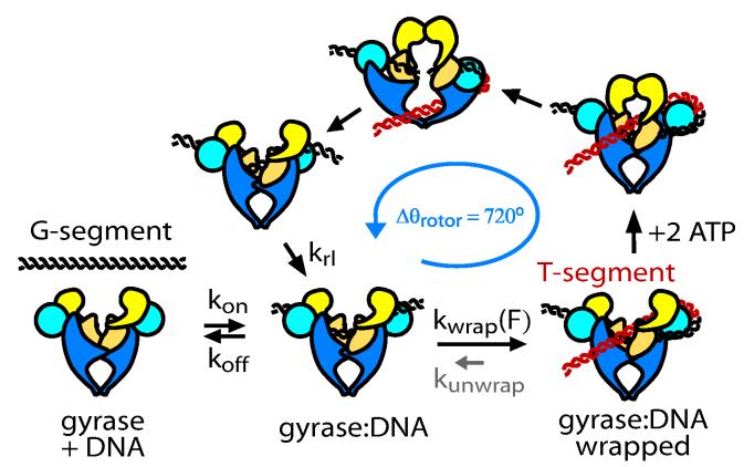
DNA gyrase is a molecular machine that uses the energy of ATP hydrolysis to introduce essential negative supercoils into DNA. The directionality of supercoiling is ensured by chiral wrapping of the DNA around a specialized domain of the enzyme before strand passage. Here we observe the activity of gyrase in real time by tracking the rotation of a submicrometre bead attached to the side of a stretched DNA molecule. In the presence of gyrase and ATP, we observe bursts of rotation corresponding to the processive, stepwise introduction of negative supercoils in strict multiples of two. Changes in DNA tension have no detectable effect on supercoiling velocity, but the enzyme becomes markedly less processive as tension is increased over a range of only a few tenths of piconewtons. This behaviour is quantitatively explained by a simple mechanochemical model in which processivity depends on a kinetic competition between dissociation and rapid, tension-sensitive DNA wrapping. In a high-resolution variant of our assay, we directly detect rotational pauses corresponding to two kinetic substeps: an ATP-independent step at the end of the reaction cycle, and an ATP-binding step in the middle of the cycle, subsequent to DNA wrapping.


Figures

Figure 1
Figure 1
Experimental design and single-molecule observations of gyrase activity. a, The molecular construct contains three distinct attachment sites and a site-specific nick, which acts as a swivel. A strong gyrase site was engineered into the lower DNA segment. b, Molecule/bead assemblies were constructed in parallel in a flow chamber and assayed with an inverted microscope equipped with permanent magnets. Each molecule was stretched between the glass coverslip and a 1 μm magnetic bead, while a 530 nm diameter fluorescent rotor bead was attached to the central biotinylated patch. In the presence of gyrase and ATP, the rotor bead underwent bursts of rotation due to the enzymatic activity of individual gyrase enzymes acting on the DNA segment below the rotor bead. c, A plot of the rotor bead angle as a function of time (averaged over a 2 second window) shows bursts of activity due to diffusional encounters of individual gyrase enzymes. The activity of the enzyme is strongly tension dependent. With the exception of the 0.35 pN trace, all traces shown were taken in the same chamber with a single concentration of gyrase, and the differences in burst density thus reflect force-dependent initiation rates. d, A histogram of the pairwise difference distribution function summed over eleven 15 - 20 minute traces (averaged over a 4 second window) at forces of 0.6 – 0.8 pN. The spacing of the peaks indicates that each catalytic cycle of the enzyme corresponds to two full rotations of the rotor bead, as expected for a type II topoisomerase such as DNA gyrase.
Figure 2
Figure 2
Modulation of gyrase activity by DNA tension. a - c, The velocity within a burst is insensitive to DNA tension, but both the processivity and initiation rate decrease rapidly as DNA tension increases (error bars, standard error of mean). c, Tension-dependent initiation rates were measured in two independent experiments after the introduction of 10 nM (green squares) or 5 nM (blue circles) gyrase.
Figure 3
Figure 3
Proposed mechanochemical model. From the state labeled gyrase:DNA there is a kinetic competition between DNA wrapping and dissociation. Wrapping is strongly inhibited by DNA tension. After wrapping and ATP binding, the enzyme is committed to a full catalytic cycle in which two negative supercoils are introduced to the DNA, causing the rotor bead to spin by Δθ = 720°. At saturating [ATP] unwrapping (small arrow labeled kunwrap) is negligible; however, see Fig. 4 b,c.
Figure 4
Figure 4
Gyrase activity observed at high resolution. a, High resolution traces (F = ∼0.8 pN) at 1 mM ATP show that the dominant pause within the catalytic cycle occurs at the two rotation mark, corresponding to either the beginning or the end of the cycle (marked by arrows). Pauses were less frequently observed in the middle of the catalytic cycle (marked by asterisks). Traces were averaged over a 300 ms window. Burst velocities in the high-resolution assay were 0.38 ± 0.04 Hz, not significantly faster than in the lower-resolution assay (0.32 ± 0.03 Hz, Fig. 2a). b, In the absence of ATP, the rotor bead angle alternates between two values, as expected for reversible DNA wrapping (black trace, F = 0.9pN). The wrapped state corresponds to a change in the angle of the rotor bead of ∼ 1.3 rotations (dashed green line), as shown (inset) by a double-Gaussian fit to the histogram of rotor bead angles for this trace. Increasing the DNA tension from 0.75 pN (green trace) to 1.3 pN (blue trace) strongly inhibits wrapping. c, Fine structure of isolated enzymatic cycles at multiple ATP concentrations. The mid-cycle pause at the ∼1 rotation mark becomes more pronounced as the ATP concentration is lowered, revealing an ATP-binding step subsequent to DNA wrapping (green traces, 1 mM ATP; blue traces, 100 μM ATP; red traces, 25 μM ATP). At 25 μM ATP, unwrapping often occurs before the cycle can be completed (final red traces).

Similar articles

Cited by

References

    1. Gellert M, Mizuuchi K, O'Dea MH, Nash HA. DNA gyrase: an enzyme that introduces superhelical turns into DNA. Proc Natl Acad Sci U S A. 1976;73:3872–6. - PMC - PubMed
    1. Champoux JJ. DNA topoisomerases: structure, function, and mechanism. Annu Rev Biochem. 2001;70:369–413. - PubMed
    1. Corbett KD, Berger JM. Structure, molecular mechanisms, and evolutionary relationships in DNA topoisomerases. Annu Rev Biophys Biomol Struct. 2004;33:95–118. - PubMed
    1. Liu LF, Wang JC. DNA-DNA gyrase complex: the wrapping of the DNA duplex outside the enzyme. Cell. 1978;15:979–84. - PubMed
    1. Liu LF, Wang JC. Micrococcus luteus DNA gyrase: active components and a model for its supercoiling of DNA. Proc Natl Acad Sci U S A. 1978;75:2098–102. - PMC - PubMed

Publication types

LinkOut - more resources