. 2006 Mar;7(3):334-40.
doi: 10.1038/sj.embor.7400615. Epub 2006 Jan 6.

Skin human papillomavirus type 38 alters p53 functions by accumulation of deltaNp73

Affiliations

Skin human papillomavirus type 38 alters p53 functions by accumulation of deltaNp73

Rosita Accardi et al. EMBO Rep. 2006 Mar.

Abstract

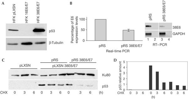
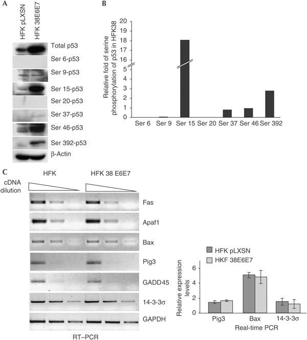
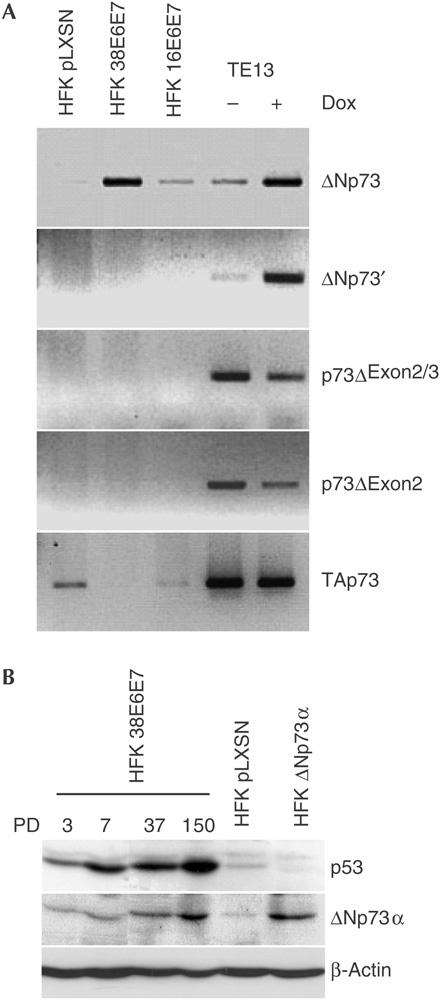
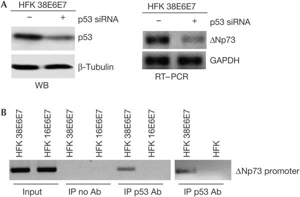
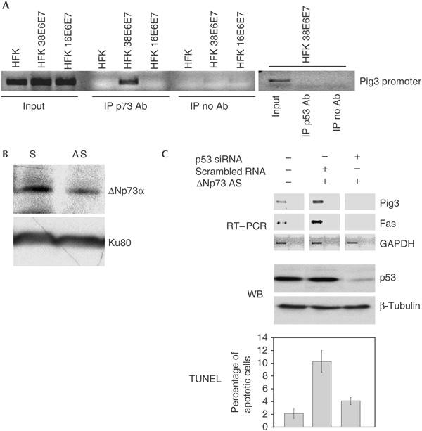
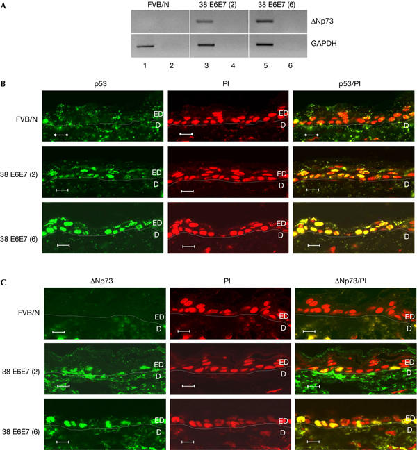
The E6 and E7 of the cutaneous human papillomavirus (HPV) type 38 immortalize primary human keratinocytes, an event normally associated with the inactivation of pathways controlled by the tumour suppressor p53. Here, we show for the first time that HPV38 alters p53 functions. Expression of HPV38 E6 and E7 in human keratinocytes or in the skin of transgenic mice induces stabilization of wild-type p53. This selectively activates the transcription of deltaNp73, an isoform of the p53-related protein p73, which in turn inhibits the capacity of p53 to induce the transcription of genes involved in growth suppression and apoptosis. DeltaNp73 downregulation by an antisense oligonucleotide leads to transcriptional re-activation of p53-regulated genes and apoptosis. Our findings illustrate a novel mechanism of the alteration of p53 function that is mediated by a cutaneous HPV type and support the role of HPV38 and deltaNp73 in human carcinogenesis.


Figures

Figure 1
Figure 1
HPV38 E6 and E7 induce stabilization of p53. (A) Human foreskin keratinocytes (HFKs) expressing HPV38 E6 and E7 harbour high levels of p53. Immunoblotting was performed using the indicated antibodies. (B) Downregulation of HPV38 E6 and E7 by small hairpin RNAs (shRNAs). HPV38-HFKs were infected with empty pRetroSuper (pRS) or pRS containing shRNA for HPV38 E6/E7 (pRS 38E6/E7) and E6 messenger RNA was determined by real-time and reverse transcription–PCR (RT–PCR). The real-time PCR data are the mean of three independent experiments. RT–PCR was performed using the indicated primers. As control, PCRs were performed on non-reverse-transcribed RNA (lanes 2,4). GAPDH, glyceraldehyde-3-phosphate dehydrogenase. (C,D) Downregulation of HPV38 E6 and E7 decreases p53 half-life. The indicated cell lines were cultured in a medium containing cycloheximide (CHX) as described in Methods. The levels of p53 and Ku80 (loading control) were determined by immunoblotting (C) and the amount of p53 and Ku80 was quantified by Fluor-S™ MultiImager (Bio-Rad) (D).
Figure 2
Figure 2
Status of p53 in HPV38-keratinocytes. (A) Characterization of p53 phosphorylation pattern. Protein extracts of the indicated cells were analysed by immunoblotting using the indicated antibodies. (B) Quantification of the increase in p53 phosphorylation in human foreskin keratinocyte (HFK) HPV38 E6/E7 in comparison with HFK pLXSN. The amounts of total and phosphorylated p53 shown in (A) were quantified as described in Methods. After normalization of the levels of the different phosphorylated forms of p53 to the signal for total p53, the relative level of p53 phosphorylation in HFK HPV38 E6/E7 cells was evaluated in comparison with primary keratinocytes (HFK pLXSN). (C) p53-regulated genes are not upregulated in HPV38-HFKs. After reverse transcription, PCR was performed on serial dilutions of the complementary DNA with the indicated primers. The real-time PCR data are the mean of three independent experiments.
Figure 3
Figure 3
HPV38 E6 and E7 expression promotes ΔNp73 accumulation. (A) The Δ isoform detected in HPV38-HFK (human foreskin keratinocyte) is expressed from the p73 p2 internal promoter. Reverse transcription–PCR (RT–PCR) was performed using specific primers for the different Δ isoforms. As control, complementary DNA of RNA extracted from untreated or doxorubicin (Dox)-treated (1 μg/μl; 24 h) p53−/− oesophageal tumour cells (TE13) was used. GAPDH, glyceraldehyde-3-phosphate dehydrogenase. (B) ΔNp73α protein is present at high levels in HPV38-keratinocytes. Protein extracts of HPV38-HFK taken at different population doublings (PD) and HFK infected with ΔNp73α retrovirus were analysed by immunoblotting.
Figure 4
Figure 4
HPV38-mediated ΔNp73 accumulation is p53 dependent. (A) ΔNp73 expression is mediated by p53. After transfection of HPV38-keratinocytes with indicated short interfering RNAs (siRNAs), p53 and ΔNp73 expression levels were determined by immunoblotting (WB, left panel) and reverse transcription–PCR (RT–PCR; right panel), respectively. GAPDH, glyceraldehyde-3-phosphate dehydrogenase; HFK, human foreskin keratinocyte. (B) p53 binds the ΔNp73 promoter in HPV38-keratinocytes. Chromatin immunoprecipitations (ChIPs) were performed on the indicated cell populations. Purified DNA from total lysate (input) and DNA co-immunoprecipitated from the control ChIP with no antibody (IP no Ab) or from the p53 immunoprecipitates (IP p53 Ab) were analysed by PCR using primers designed within the p73 p2 internal promoter.
Figure 5
Figure 5
Downregulation of ΔNp73 leads to re-activation of p53-regulated genes and apoptosis. (A) Pig3 promoter is bound to ΔNp73, but not to p53, in HPV38-keratinocytes. Chromatin immunoprecipitations were performed as described in the legend of Fig 3C and Methods. p53 and p73 immunoprecipitates were analysed by PCR using Pig3 promoter primers. HFK, human foreskin keratinocyte. (B) Antisense oligonucleotide reduces ΔNp73 protein levels. ΔNp73 antisense oligonucleotides were transfected into HPV38- keratinocytes. ΔNp73α and Ku80 levels were detected by immunoblotting. AS, antisense; S, sense. (C) ΔNp73 downregulation induces p53-mediated apoptosis. HPV38-keratinocytes were transfected with different oligonucleotides as indicated. PCR was performed on complementary DNAs using indicated primers (reverse transcription–PCR (RT–PCR), top panel). Control PCR was performed using non-reverse transcribed RNA as template (far right of each panel). The p53 levels were determined by immunoblotting (WB, middle panel). Apoptosis was detected by TdT-mediated dUTP nick end labelling (TUNEL) assay (bottom panel). GAPDH, glyceraldehyde-3-phosphate dehydrogenase; siRNA, small interfering RNA.
Figure 6
Figure 6
p53 and ΔNp73 accumulate in keratinocytes of HPV38 E6/E7 transgenic mice. (A) ΔNp73 messenger RNA is expressed in the epidermis of HPV38 E6/E7 transgenic mice. PCR was performed on complementary DNA prepared from total RNA of non-transgenic and transgenic mice. As control, PCR was performed on non-reverse-transcribed RNA (lanes 2,4,6). GAPDH, glyceraldehyde-3-phosphate dehydrogenase. (B,C) Accumulation of p53 and ΔNp73 in skin keratinocytes of HPV38 E6/E7 transgenic mice. Skin sections were prepared and stained with antibodies against p53 (C) or p73 (D) (green) and propidium iodide (PI, red) as described in Methods. Original photographs were taken at × 63 magnification. The derma (D) and epidermis (ED) are indicated. Scale bars, 10 μm (B,C).

Similar articles

Cited by

References

    1. Allart S, Martin H, Detraves C, Terrasson J, Caput D, Davrinche C (2002) Human cytomegalovirus induces drug resistance and alteration of programmed cell death by accumulation of ΔN-p73α. J Biol Chem 277: 29063–29068 - PubMed
    1. Auewarakul P, Gissmann L, Cidarregui A (1994) Targeted expression of the E6 and E7 oncogenes of human papillomavirus type 16 in the epidermis of transgenic mice elicits generalized epidermal hyperplasia involving autocrine factors. Mol Cell Biol 14: 8250–8258 - PMC - PubMed
    1. Bode AM, Dong Z (2004) Post-translational modification of p53 in tumorigenesis. Nat Rev Cancer 4: 793–805 - PubMed
    1. Caldeira S, Zehbe I, Accardi R, Malanchi I, Dong W, Giarre M, deVilliers E-M, Filotico R, Boukamp P, Tommasino M (2003) The E6 and E7 proteins of cutaneous human papillomavirus type 38 display transforming properties. J Virol 77: 2195–2206 - PMC - PubMed
    1. Caldeira S, Filotico R, Accardi R, Zehbe I, Franceschi S, Tommasino M (2004) p53 mutations are common in human papillomavirus type 38-positive non-melanoma skin cancers. Cancer Lett 209: 119–124 - PubMed

Publication types

MeSH terms