Comparative Study
. 2006 Jan 4;26(1):210-6.
doi: 10.1523/JNEUROSCI.4307-05.2006.

Multivesicular release at Schaffer collateral-CA1 hippocampal synapses

Affiliations
Comparative Study

Multivesicular release at Schaffer collateral-CA1 hippocampal synapses

Jason M Christie et al. J Neurosci. .

Abstract

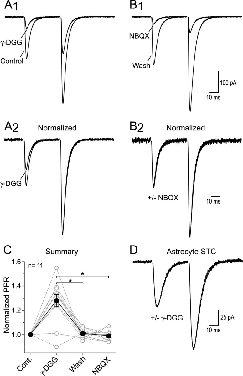
Whether an individual synapse releases single or multiple vesicles of transmitter per action potential is contentious and probably depends on the type of synapse. One possibility is that multivesicular release (MVR) is determined by the instantaneous release probability (Pr) and therefore can be controlled by activity-dependent changes in Pr. We investigated transmitter release across a range of Pr at synapses between Schaffer collaterals (SCs) and CA1 pyramidal cells in acute hippocampal slices using patch-clamp recordings. The size of the synaptic glutamate transient was estimated by the degree of inhibition of AMPA receptor EPSCs with the rapidly equilibrating antagonist gamma-D-glutamylglycine. The glutamate transient sensed by AMPA receptors depended on Pr but not spillover, indicating that multiple vesicles are essentially simultaneously released from the same presynaptic active zone. Consistent with an enhanced glutamate transient, increasing Pr prolonged NMDA receptor EPSCs when glutamate transporters were inhibited. We suggest that MVR occurs at SC-CA1 synapses when Pr is elevated by facilitation and that MVR may be a phenomenon common to many synapses throughout the CNS.


Figures

Figure 1.
Figure 1.
Paired-pulse EPSCs are differentially blocked by γ-DGG. A1, AMPA receptor-mediated EPSCs evoked by paired stimuli (40 ms) in control and in γ-DGG (2 mm) are superimposed. A2, Traces from A1 are normalized to the peak of the second EPSC. B1, Paired EPSCs after γ-DGG washout and in NBQX (400 nm). B2, Traces from B1, normalized to the peak of the second EPSC. C, Summary data show that PPR increases in the presence of γ-DGG. White circles represent the normalized PPR (see Materials and Methods) of individual experiments, and black circles are the mean values. Error bars indicate ±SEM. D, STCs from area CA1 astrocytes are unaffected by γ-DGG (2 mm). All traces are the average of multiple responses.
Figure 2.
Figure 2.
Effects of the glutamate uptake antagonist TBOA. A, STCs recorded in a CA1 astrocyte. Responses in control and in TBOA (10 μm) are superimposed. B, NMDA receptor-mediated EPSCs from a CA1 pyramidal cell in control and in the presence of TBOA(10μm). Inset, The normalized EPSCs are shown expandedin time. C, AMPA receptor-mediated EPSCs in control and in TBOA (10 μm). D, Summary data show the effects of TBOA on the amplitude and decay times constants of AMPA and NMDA receptor-mediated EPSCs and astrocyte STCs. Experiments were repeated in the presence of the broad-spectrum metabotropic glutamate receptor antagonist LY341495.
Figure 3.
Figure 3.
γ-DGG inhibition is unaffected by glutamate uptake antagonism. A1, AMPA receptor-mediated EPSCs in control and in γ-DGG (2 mm). A2, Responses are normalized to the peak of EPSC2. B1, EPSCs in TBOA (10μm) and in TBOA plus γ-DGG (2 mm). B2, Normalized EPSCs show the differential block by γ-DGG. For comparison, dashed lines indicate the normalized peaks of EPSC1 in control and γ-DGG. C, Summary data show that the γ-DGG-dependent increase in the PPR was unaltered by uptake block. D, Plot of γ-DGG inhibition of EPSC1 (left) and EPSC2 (right) in the absence and presence of TBOA (10 μm).
Figure 4.
Figure 4.
γ-DGG inhibition depends on release probability. A, AMPA receptor-mediated EPSCs evoked in three Ca+2e conditions from three different cells. Responses from the same cell show inhibition by γ-DGG(2mm) or NBQX (400 nm) on the top and bottom, respectively. For comparison, dashed lines indicate the amplitude of the inhibited EPSC in 1.5 mm Ca+2e.B1, Summary data show that γ-DGG inhibition was dependent on the Ca+2e B2, NBQX inhibited EPSCs to the same extent. C1, AMPA receptor-mediated EPSCs evoked in 1.25 mmCa+2e in control and in γ-DGG (2mm). C2, Traces from C1 normalized to the peak of the second EPSC.C3, Summary plot shows that the PPR was unaltered by γ-DGG. D1, EPSCs evoked in 2.5 mm Ca+2e in control and in γ-DGG (2mm).D2, Traces from D1 normalized to the peak of the second EPSC. D3, Summary plot shows thatγ-DGG, but not NBQX (400 nm), increased PPR.
Figure 5.
Figure 5.
MVR can enhance the magnitude of glutamate spillover. A1, NMDA receptor-mediated EPSCs evoked in 1.5 mm Ca+2e in control and in TBOA (10 μm). Inset, Normalized EPSCs expanded in time. A2, EPSCs evoked after Ca+2e were elevated to 2.5 mm. Responses in control and in TBOA (10 μm) are shown super imposed. The stimulation intensity was reduced so that the control EPSC in 2.5 mm Ca+2e approximately matched the control EPSC evoked in 1.5 mm Ca+2e. Inset, Normalized responses are expanded in time. B1, Summary data show that TBOA increased the decay time constants (τ1 and τ2, left and right, respectively) of EPSCs to agreater extent in 2.5 mm Ca+2e. B2, The TBOA-dependent increase of the EPSC integral was also larger in 2.5 mm Ca+2e (left). The EPSC peak amplitude was enhanced to a similar extent (right). C, The control EPSCs in 1.5 and 2.5 mm Ca+2e are normalized and superimposed.

Similar articles

Cited by

References

    1. Arnth-Jensen N, Jabaudon D, Scanziani M (2002) Cooperation between independent hippocampal synapses is controlled by glutamate uptake. Nat Neurosci 5: 325–331. - PubMed
    1. Asztely F, Erdemli G, Kullmann D (1997) Extrasynaptic glutamate spillover in the hippocampus: the role of active glutamate uptake. Neuron 18: 281–293. - PubMed
    1. Auger C, Kondo S, Marty A (1998) Multivesicular release at single functional synaptic sites in cerebellar stellate and basket cells. J Neurosci 18: 4532–4547. - PMC - PubMed
    1. Barbour B, Häusser M (1997) Intersynaptic diffusion of neurotransmitter. Trends Neurosci 20: 377–384. - PubMed
    1. Bergles DE, Jahr CE (1997) Synaptic activation of glutamate transporters in hippocampal astrocytes. Neuron 19: 1297–1308. - PubMed

Publication types

Substances