. 2006 Jan;168(1):104-14; quiz 363.
doi: 10.2353/ajpath.2006.050575.

Chronic psychological stress in rats induces intestinal sensitization to luminal antigens

Affiliations

Chronic psychological stress in rats induces intestinal sensitization to luminal antigens

Ping-Chang Yang et al. Am J Pathol. 2006 Jan.

Abstract

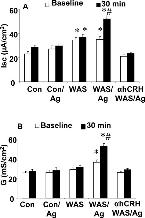
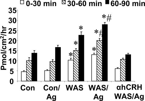
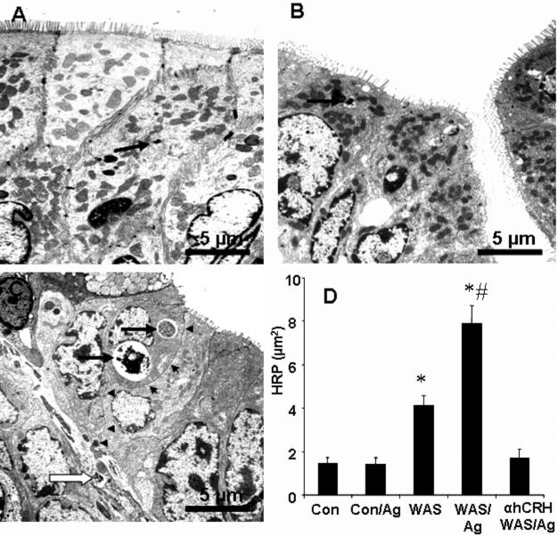
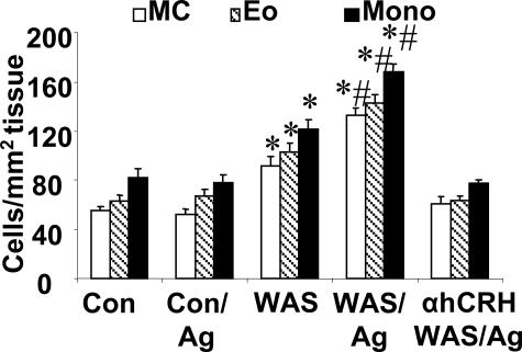
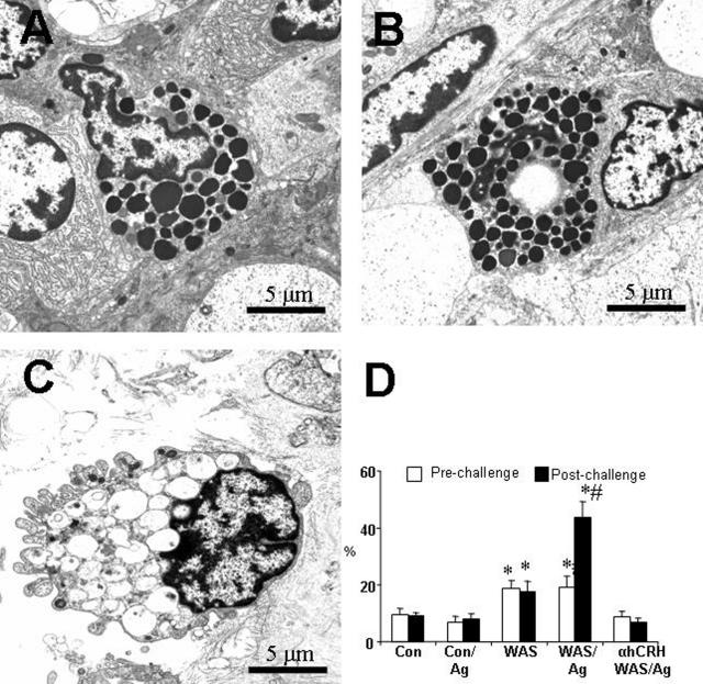
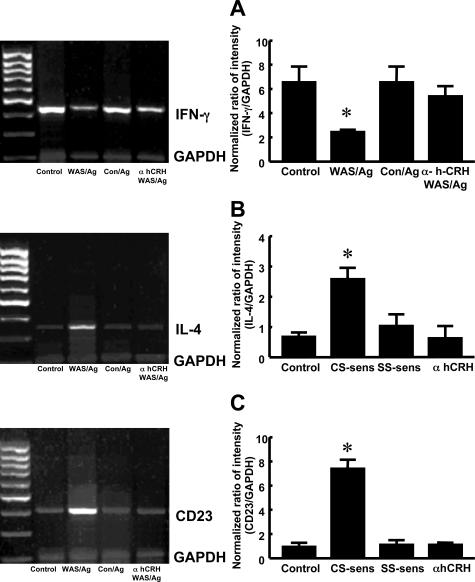
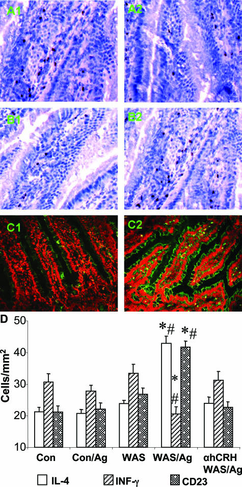
There is increasing evidence that stress plays a role in the pathophysiology of chronic intestinal disorders, but the mechanisms remain unclear. Previous studies in rats have revealed that stress decreases gut barrier function and allows excessive uptake of luminal material. Here, we investigated whether chronic psychological stress acts to induce sensitization of intestinal tissues to oral antigens. Rats were subjected to 1 hour per day of water avoidance stress or sham stress daily for 10 days, and horseradish peroxidase (HRP) was delivered by gavage on day 5. Studies to determine sensitization were conducted on day 20. All stressed rats developed HRP-specific IgE antibodies, antigen-induced intestinal secretion, and increased numbers of inflammatory cells in gut mucosa. Luminal HRP was absorbed more readily by enterocytes of stressed animals. In addition, stressed rats had increased expression of interleukin-4 and decreased expression of interferon-gamma in gut mucosa, a cytokine profile that is typical of allergic conditions. Treatment of stressed rats with an antagonist to corticotropin-releasing hormone (previously shown to inhibit stress-enhanced gut permeability) eliminated the manifestations of intestinal hypersensitivity. Our results indicate that the presence of oral antigen during chronic psychological stress alters the immune response (to sensitization rather than oral tolerance) and causes subsequent antigen-induced gut pathophysiology.


Figures

Figure 1
Figure 1
Electrophysiological responses of jejunal tissues to antigen challenge. Ussing chambers were used for the study. Jejunal tissues were obtained from naive control rats (Con), stressed (WAS/Ag) rats, sham-stressed (Con/Ag) rats, WAS-only (WAS) rats, and WAS rats pretreated with the CRH antagonist, α-hCRH (αhCRH/WAS/Ag). All rats except Con group received a bolus of intragastric HRP at day 5 of the stress/sham protocol; 15 days later tissues were studied in Ussing chambers. A: Secretory response. Isc was recorded at baseline and 30 minutes after adding HRP antigen to the mucosal (apical) buffer. B: Conductance. G was recorded at baseline and 30 minutes after adding HRP antigen to the apical buffer. Values indicate mean ± SEM; n = 6 rats in each group (four tissues per rat averaged to obtain each rat value).
Figure 2
Figure 2
Effect of sensitization and Ag challenge on mucosal to serosal flux of HRP. Flux of HRP was determined across the jejunal epithelium that was obtained from naive control rats (Con), stressed (WAS/Ag) rats, sham-stressed (Con/Ag) rats, WAS-only (WAS) rats, and WAS rats pretreated with the CRH antagonist, α-hCRH (αhCRH/WAS/Ag). HRP was measured in serosal samples after addition of 10−5 mol/L HRP to the luminal buffer, and fluxes were calculated for three flux periods: 0 to 30 minutes, 30 to 60 minutes, and 60 to 90 minutes. Values are expressed as means ± SE, pmol/cm2/hour; n = 4 rats/group; *P < 0.05, compared with Con rats; #P < 0.05 compared with values in WAS-only rats in each period.
Figure 3
Figure 3
Pathways of transepithelial transport of HRP. Jejunal tissues were obtained from naive control rats (Con), stressed (WAS/Ag) rats, sham-stressed (Con/Ag) rats, WAS-only (WAS) rats, and WAS rats pretreated with the CRH antagonist, α-hCRH (αhCRH/WAS/Ag). All rats except naive controls received a bolus of intragastric HRP at day 5 of the stress/sham protocol; 15 days later the tissues were obtained and processed for Ussing chamber study and electron microscopy. A–C: Electron photomicrographs show endosomes containing HRP (arrows) within enterocytes. A: Representative section from a Con/Ag rat. B: Representative section from a WAS/Ag/α-hCRH-treated rat. C: A representative section from a WAS/Ag rat shows HRP endosomes (arrows) in the epithelial cells, HRP products in the paracellular spaces (arrowheads), and HRP endosome in the lamina propria (open arrow). D: Image analysis of the total area of HRP-containing endosomes within a fixed sized window (300 μm2) in enterocytes. Values indicate mean ± SEM; n = 6 rats in each group (12 views per rat section averaged to obtain each rat value).
Figure 4
Figure 4
Inflammatory cells in gut mucosa. Numbers of mast cells (MC), eosinophils (Eo), and mononuclear cells (Mono) were counted in coded sections. Jejunal tissues were obtained from naive control rats (Con), stressed (WAS/Ag) rats, sham-stressed (Con/Ag) rats, WAS-only (WAS) rats, and WAS rats pretreated with the CRH antagonist, α-hCRH (αhCRH/WAS/Ag). Tissues were fixed and stained as indicated in Materials and Methods. The results are expressed as number of cells per mm2. Bars indicate means ± SEM; values were obtained from 25 fields per rat (150 per group). *P < 0.05, compared with naïve controls. #P < 0.05, compared with the rats of WAS group.
Figure 5
Figure 5
Mast cell activation. Jejunal tissues were obtained from naive control rats (Con), stressed (WAS/Ag) rats, sham-stressed (Con/Ag) rats, WAS-only (WAS) rats, and WAS rats pretreated with the CRH antagonist, α-hCRH (αhCRH/WAS/Ag). All rats except naive controls received a bolus of intragastric HRP at day 5 of the stress/sham protocol; 15 days later tissues were obtained and mounted in Ussing chambers. Ninety minutes after HRP antigen challenge, tissues were removed and processed for electron microscopy. A–C: Electron photomicrographs show mucosal mast cells in the lamina propria. A and B: Representative sections from a Con/Ag rat (A) and rat treated with α-hCRH (B) show normal mast cells with electron dense granules. C: Representative section from a WAS/Ag rat shows a mast cell with depleted granules. D: Percentage of mast cells degranulated after HRP antigen challenge as measured by empty granules or reduced density of granules. Values indicate mean ± SEM; n = 6 rats in each group (20 random views per rat section averaged to obtain each rat value). *P < 0.05, compared with Con group; #P < 0.05, compared with WAS-only group.
Figure 6
Figure 6
Message and protein expression of INF-γ, IL-4, and CD23. Jejunal tissues were obtained from naive control rats, stressed (WAS/Ag) rats, sham-stressed (Con/Ag) rats, and WAS/Ag rats pretreated with the CRH antagonist, α-hCRH. All rats except naive controls received a bolus of intragastric HRP at day 5 of the stress/sham protocol; 15 days later tissues were obtained. A: mRNA was purified from jejunal mucosa. Reverse transcriptase-PCR was used to determine mRNA (primers as in Table 1); the ratio of intensity of mRNA bands was normalized to GAPDH. Values indicate mean ± SEM; n = 6 rats in each group. B: Numbers of positively stained cells (per mm2) in the lamina propria (for IFN-γ and IL-4) or epithelium (for CD23). Values indicate mean ± SEM; n = 6 rats in each group (10 random fields per rat averaged to obtain each rat value).
Figure 7
Figure 7
Immunohistochemistry of jejunal mucosa showing protein expression of IFN-γ, IL-4, and CD23. Representative sections include tissues from Con/Ag rats (A1–C1) and WAS/Ag rats (A2–C2). A: Representative section showing cells in the lamina propria stained for IFN-γ. B: Representative section showing cells in the lamina propria stained for IL-4. C: Representative section showing epithelial cells and cells in the lamina propria stained for CD23. D: Counts of positive stained cells in jejunal tissues that were obtained from naive control rats (Con), stressed (WAS/Ag) rats, sham-stressed (Con/Ag) rats, WAS-only (WAS) rats, and WAS rats pretreated with the CRH antagonist, α-hCRH (αhCRH/WAS/Ag). The results are expressed as number of cells per mm2. Bars indicate means ± SEM; values were obtained from 25 fields per rat (150 per group). *P < 0.05, compared with naïve controls. #P < 0.05, compared with the rats of WAS group.

Comment in

Similar articles

Cited by

References

    1. Collins SM. Stress and the gastrointestinal tract IV. Modulation of intestinal inflammation by stress: basic mechanisms and clinical relevance. Am J Physiol. 2001;280:G315–G318. - PubMed
    1. Monnikes H, Tebbe JJ, Hildebrandt M, Arck P, Osmanoglou E, Rose M, Klapp B, Wiedenmann B, Heymann-Monnikes I. Role of stress in functional gastrointestinal disorders. Evidence for stress-induced alterations in gastrointestinal motility and sensitivity. Dig Dis. 2001;19:201–211. - PubMed
    1. Hart A, Kamm MA. Mechanisms of initiation and perpetuation of gut inflammation by stress. Aliment Pharmacol Ther. 2002;16:2017–2028. - PubMed
    1. Kelsay K. Psychological aspects of food allergy. Curr Allergy Asthma Rep. 2003;3:41–46. - PubMed
    1. Guarner F, Casellas F, Borruel N, Antolin M, Videla S, Vilaseca J, Malagelada JR. Role of microecology in chronic inflammatory bowel diseases. Eur J Clin Nutr. 2002;56:S34–S38. - PubMed

Publication types

MeSH terms