

Influenza A virus (H5N1) infection in cats causes systemic disease with potential novel routes of virus spread within and between hosts
- PMID: 16400021
- PMCID: PMC1592682
- DOI: 10.2353/ajpath.2006.050466
Influenza A virus (H5N1) infection in cats causes systemic disease with potential novel routes of virus spread within and between hosts
Abstract
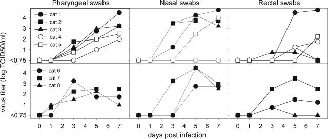
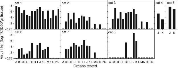
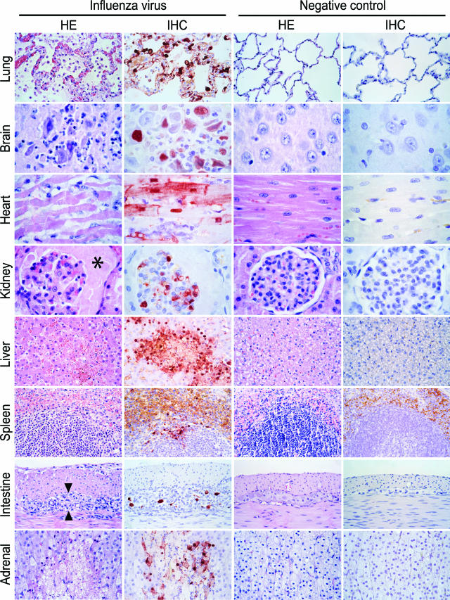
The ongoing outbreak of avian influenza A virus (subtype H5N1) infection in Asia is of great concern because of the high human case fatality rate and the threat of a new influenza pandemic. Case reports in humans and felids suggest that this virus may have a different tissue tropism from other influenza viruses, which are normally restricted to the respiratory tract in mammals. To study its pathogenesis in a mammalian host, domestic cats were inoculated with H5N1 virus intratracheally (n = 3), by feeding on virus-infected chicks (n = 3), or by horizontal transmission (n = 2) and examined by virological and pathological assays. In all cats, virus replicated not only in the respiratory tract but also in multiple extra-respiratory tissues. Virus antigen expression in these tissues was associated with severe necrosis and inflammation 7 days after inoculation. In cats fed on virus-infected chicks only, virus-associated ganglioneuritis also occurred in the submucosal and myenteric plexi of the small intestine, suggesting direct infection from the intestinal lumen. All cats excreted virus not only via the respiratory tract but also via the digestive tract. This study in cats demonstrates that H5N1 virus infection causes systemic disease and spreads by potentially novel routes within and between mammalian hosts.
Figures
Comment in
-
Avian influenza: Virchow's reminder.Am J Pathol. 2006 Jan;168(1):6-8. doi: 10.2353/ajpath.2006.050979. Am J Pathol. 2006. PMID: 16400004 Free PMC article. Review. No abstract available.
References
-
- Li KS, Guan Y, Wang J, Smith GJ, Xu KM, Duan L, Rahardjo AP, Puthavathana P, Buranathai C, Nguyen TD, Estoepangestie AT, Chaisingh A, Auewarakul P, Long HT, Hanh NT, Webby RJ, Poon LL, Chen H, Shortridge KF, Yuen KY, Webster RG, Peiris JS. Genesis of a highly pathogenic and potentially pandemic H5N1 influenza virus in eastern Asia. Nature. 2004;430:209–213. - PubMed
-
- Tran TH, Nguyen TL, Nguyen TD, Luong TS, Pham PM, Nguyen VC, Pham TS, Vo CD, Le TQ, Ngo TT, Dao BK, Le PP, Nguyen TT, Hoang TL, Cao VT, Le TG, Nguyen DT, Le HN, Nguyen KT, Le HS, Le VT, Christiane D, Tran TT, Menno de J, Schultsz C, Cheng P, Lim W, Horby P, Farrar J. Avian influenza A (H5N1) in 10 patients in Vietnam. N Engl J Med. 2004;350:1179–1188. - PubMed
-
- Keawcharoen J, Oraveerakul K, Kuiken T, Fouchier RA, Amonsin A, Payungporn S, Noppornpanth S, Wattanodorn S, Theambooniers A, Tantilertcharoen R, Pattanarangsan R, Arya N, Ratanakorn P, Osterhaus DM, Poovorawan Y. Avian influenza H5N1 in tigers and leopards. Emerg Infect Dis. 2004;10:2189–2191. - PMC - PubMed
-
- Kuiken T, Rimmelzwaan G, Van Riel D, Van Amerongen G, Baars M, Fouchier R, Osterhaus A. Avian H5N1 influenza in cats. Science. 2004;306:241. - PubMed
Publication types
MeSH terms
LinkOut - more resources
Full Text Sources
Other Literature Sources
Medical
Miscellaneous
