. 2006 Jan;168(1):292-309.
doi: 10.2353/ajpath.2006.050429.

Caveolin-1 deficiency (-/-) conveys premalignant alterations in mammary epithelia, with abnormal lumen formation, growth factor independence, and cell invasiveness

Affiliations

Caveolin-1 deficiency (-/-) conveys premalignant alterations in mammary epithelia, with abnormal lumen formation, growth factor independence, and cell invasiveness

Federica Sotgia et al. Am J Pathol. 2006 Jan.

Abstract

During breast cancer development, the luminal space of the mammary acinar unit fills with proliferating epithelial cells that exhibit growth factor-independence, cell attachment defects, and a more invasive fibroblastic phenotype. Here, we used primary cultures of mammary epithelial cells derived from genetically engineered mice to identify caveolin-1 (Cav-1) as a critical factor for maintaining the normal architecture of the mammary acinar unit. Isolated cultures of normal mammary epithelial cells retained the capacity to generate mammary acini within extracellular matrix. However, those from Cav-1 (-/-) mice exhibited defects in three-dimensional acinar architecture, including disrupted lumen formation and epidermal growth factor-independent growth due to hyperactivation of the p42/44 mitogen-activated protein kinase cascade. In addition, Cav-1-null mammary epithelial cells deprived of exogenous extracellular matrix underwent a spontaneous epithelial-mesenchymal transition, with reorganization of the actin cytoskeleton, and E-cadherin redistribution. Mechanistically, these phenotypic changes appear to be caused by increases in matrix metalloproteinase-2/9 secretion and transforming growth factor-beta/Smad-2 hyperactivation. Finally, loss of Cav-1 potentiated the ability of growth factors (hepatocyte growth factor and basic fibroblast growth factor) to induce mammary acini branching, indicative of a more invasive fibroblastic phenotype. Thus, a Cav-1 deficiency profoundly affects mammary epithelia by modulating the activation state of important signaling cascades. Primary cultures of Cav-1-deficient mammary epithelia will provide a valuable new model to study the spatial/temporal progression of mammary cell transformation.


Figures
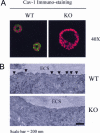
Figure 1
Figure 1
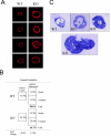
Cav-1-deficient mammary epithelial cells form three-dimensional acini-like structures but are devoid of caveolae. Primary mammary epithelial cells derived from WT and Cav-1-null mice were grown embedded in Matrigel. Under these conditions, both WT and Cav-1 KO mammary epithelial cells develop three-dimensional epithelial structures that closely resemble mammary gland acini. A: Day 8 WT and Cav-1-deficient acini were immunostained with anti-Cav-1 IgG and counterstained with propidium iodide to visualize cell nuclei. Single optical sections were acquired with a Bio-Rad Radiance 2000 scanning laser confocal microscope. Note that Cav-1 is properly expressed in WT mammary epithelial cells but is clearly absent in Cav-1 KO acini. B: Electron microscopic analysis of day 20 acini reveals that loss of Cav-1 disrupts caveolae formation in mammary epithelial cells. Note the presence of several caveolae, defined as 50- to 100-nm plasma membrane invaginations, in WT acini (arrows). On the contrary, Cav-1-deficient acini exhibit a complete loss of caveolae. ECS, extracellular space. Scale bar, 200 nm.
Figure 2
Figure 2
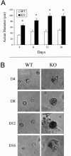
Cav-1 KO mammary epithelial cells form dramatically larger acini. To form acini-like structures, WT and Cav-1-null mammary epithelial cells were grown in reconstituted basement membrane, ie, Matrigel. Note that loss of Cav-1 confers a growth advantage. A: Growth curves of WT and Cav-1 KO acini. Acinar diameter was measured at days 4, 8, 12, and 16, with the support of Image J software. Note that, at any given time point, Cav-1-null acini demonstrate an ∼2- to 2.2-fold increase in size. In addition, although WT acini reach a growth-arrested state by day 8, Cav-1-deficient acini continue to enlarge and do not undergo growth arrest until day 12. More than 50 acini for each genotype were scored at a given time point. *P ≤ 0.0002. Error bars, SEM. B: Phase micrographs of WT and Cav-1 KO acini. Note that WT mammary epithelial cells develop acini-like structures with a regular shape, whereas Cav-1-null acini appear larger and morphologically distinct. Representative examples at days 4, 8, 12, and 16 are shown.
Figure 3
Figure 3
Cav-1 KO mammary acini show thickened walls with abnormal three-dimensional lumen formation. WT and Cav-1-null mammary epithelial cells were grown in reconstituted basement membrane, ie, Matrigel, to generate mammary acini. Interestingly, note that loss of Cav-1 results in wall thickening and abnormal lumen formation. A: Nuclear staining. Day 8 WT and Cav-1 KO acini were stained with propidium iodide to visualize nuclei (red) and appreciate lumen formation. WT acini exhibit a single layer of mammary epithelial cells lining a hollow lumen. Note that loss of Cav-1 induces wall thickening, to two or more cell layers, and in certain cases elicits luminal space filling. B: Quantitation of luminal obstruction. WT and Cav-1-null acini were stained with propidium iodide to visualize lumen formation. Acini were then scored based on the presence of a hollow lumen or for partial or complete luminal filling. Partial luminal obstruction was defined as mammary acini that were more than one cell layer in thickness. Note that ∼70% of WT acini contain a hollow lumen. In contrast, Cav-1-null acini displayed an ∼2.3-fold increase in luminal filling (partial and complete); similarly, partial luminal filling was increased by approximately threefold. Conversely, the percentage of Cav-1-deficient acini with a hollow lumen is reduced by ∼50%, as compared with WT acini. More than 150 acini were scored for each genotype. C: Light microscopy. Thin sections of day 20 WT and Cav-1 KO acini were cut and stained with toluidine blue. Images were acquired at low magnification, and montages were assembled to illustrate the wall thickening of Cav-1-deficient acini. Note that, in certain instances, the lumen (L) appears completely filled. Original magnifications, ×40 (A).
Figure 4
Figure 4
Cav-1-null acini show hyperactivation of the p42/44-MAP kinase signaling cascade, and EGF-independent growth. A: Hyperactivation of the Ras-p42/44-MAP kinase pathway in Cav-1 KO acini. Lysates from day 18 WT and Cav-1-deficient acini were subjected to Western blot analysis with antibodies against the activated phosphorylated form of ERK-1/2. Note that the Ras-p42/44-MAP kinase signaling cascade is hyperactivated in Cav-1-null acini. Equal loading was assessed by Western blot with a control phospho-independent antibody that recognizes total ERK-1/2. Immunoblotting with E-cadherin is shown as an additional control for equal epithelial protein content. B–D: EGF-independent growth of Cav-1-null acini. To evaluate whether loss of Cav-1 imparts growth factor independence, WT and Cav-1 KO three-dimensional epithelial structures were cultured either in the absence or in the presence of EGF, for up to 12 days. Images were acquired at days 4, 8, and 12. B: Phase images of EGF-deprived acini. In the absence of EGF, WT acini are very small and unable to grow. On the contrary, Cav-1-null mammary epithelial cells retain the ability to proliferate and to form acini-like spheroids without EGF. C: Acinar growth: size. The diameter of WT and Cav-1 KO acini, cultured with or without EGF, was measured at days 4, 8, and 12. At least 50 acini for each genotype were scored at any given time point. As expected, in the presence of EGF, Cav-1-null acini display an obvious delay in reaching a growth-arrested state (day 12 versus day 8 of WT acini), and exhibit an approximately twofold size increase, as compared to WT acini. However, in the absence of EGF, WT mammary epithelial cells develop as very small spheroids, which do not enlarge, suggesting that EGF is normally required for the growth and proliferation of three-dimensional cultures of mammary epithelial cells. In striking contrast, Cav-1-deficient acini cultured without EGF grow at a comparable rate as when they are cultured in the presence of EGF. More than 50 acini for each genotype were scored at a given time point. *P ≤ 0.00004. Error bars, SEM. D: Acinar growth: number of acini per high-power field. WT and Cav-1 KO acini were cultured with or without EGF in parallel experiments. The number of acini per high-power field was counted at days 4, 8, and 12. At least 20 high-power fields were scored for each genotype at any given time point. Note that, when cultured with EGF, WT and Cav-1 KO mammary epithelial cells form a similar number of acini, demonstrating that we seeded a comparable amount of WT and Cav-1-null cells into the wells. However, parallel experiments performed in the absence of EGF, show that the number of WT, but not that of Cav-1 KO acini, is greatly affected by a lack of EGF. Remarkably, in the absence of EGF, the number of Cav-1 KO acini is approximately fourfold increased, as compared to WT acini. *P ≤ 0.00018. Error bars, SEM.
Figure 5
Figure 5
Cav-1-null organoids show impaired attachment/spreading on glass. Intact mammary acini, ie, organoids, were purified from WT and Cav-1 KO mice and seeded onto glass coverslips. Note that after 4 days in culture, WT organoids were able to attach and grow out as a mammary epithelial cell monolayer. On the contrary, at the same 4-day time point, Cav-1 KO organoids attached but did not spread out into a cell monolayer. However, after 7 to 8 days in culture, both WT and Cav-1 KO organoids were able to grow as a mammary epithelial cell monolayer on glass (not shown). Original magnifications, ×10.
Figure 6
Figure 6
Cav-1 KO mammary epithelial cells display increased expression and secretion of MMP-9 and MMP-2. A and B: WT and Cav-1-deficient organoids, ie, intact mammary acini, were seeded onto chamber slides, cultured for 8 to 9 days, and allowed to spread as a monolayer of mammary epithelial cells. Cells were then immunostained with antibodies against MMP-9 (A) and MMP-2 (B). Note that both MMP-9 and MMP-2 expression levels are dramatically increased in Cav-1-null mammary epithelial cells. Note the strong staining within the extracellular matrix and intracellularly. C: Western blot analysis of day 18 WT and Cav-1-null acini was performed with antibodies against MMP-9 and MMP-2. Note that MMP-9 and MMP-2 expression levels are increased in Cav-1 KO mammary epithelial cells, as compared to their WT counterparts. Immunoblotting with E-cadherin is also shown as a control for equal epithelial protein content. Regarding the apparent molecular weight of MMP-2, the band that we observe is ∼66 kd, which is more consistent with the activated form. D: Increased MMP-9 secretion in Cav-1 KO acini. Media from WT and Cav-1-null acini was collected at days 12, 16, and 20 and concentrated fourfold using a centrifugal filter device. Equal amounts of concentrated media were resolved by SDS-PAGE, followed by Western blot analysis with antibodies against MMP-9. Note that Cav-1-deficient acini secrete higher levels of MMP-9 than WT acini, at any given time point. It is important to note that these experiments were performed with medium containing reduced serum content (2% horse serum; see Materials and Methods). However, the mobility of MMP-9 may still be shifted upward because of the presence of serum albumin.
Figure 7
Figure 7
Loss of type IV collagen in Cav-1-null organoids. Intact mammary acini, organoids, isolated from WT and Cav-1 KO mammary glands were cultured in Matrigel for 5 days and immunostained with a monoclonal antibody that recognizes type IV collagen (green). Nuclei were counterstained with propidium iodide (red). Note that WT organoids exhibit the basal deposition of extracellular matrix components, such as collagen type IV. On the contrary, Cav-1 KO organoids display a loss of collagen IV, probably because of the increased secretion of MMPs, which possess collagenase activity.
Figure 8
Figure 8
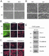
Cav-1-deficient mammary epithelial cells undergo a spontaneous epithelial mesenchymal transition (EMT). Organoids from WT and Cav-1 KO mammary glands were seeded onto glass chamber slides. During prolonged culture on glass, Cav-1-null mammary epithelial cells underwent spontaneous morphological changes, resembling an epithelial mesenchymal transition (EMT). A: Phase images. Note that after 7 and 9 days in culture, WT mammary epithelial cells retained a clear epithelial phenotype. On the contrary, at the same time points, Cav-1 KO mammary epithelial cells lost their epithelial morphology and acquired a more fibroblastic shape. B: Immunofluorescence with phalloidin, E-cadherin, and β-catenin on WT and Cav-1 KO mammary epithelial cells demonstrates that Cav-1 KO mammary epithelial cells undergo profound morphological changes. Phalloidin staining reveals that actin is no longer membrane-bound but rather is organized in stress fibers. The distribution of E-cadherin and β-catenin is profoundly altered in Cav-1 KO mammary epithelial cells. In WT cells, E-cadherin and β-catenin are localized to the plasma membrane, at areas of cell-cell contact. On the contrary, in Cav-1-null cells, E-cadherin is redistributed to an intracellular compartment, whereas β-catenin staining reveals a ruffled or jagged-edge membrane morphology. C: Immunofluorescence with cytokeratin 5/8 and 18 on WT and Cav-1 KO mammary epithelial cells shows an abundance of epithelial-specific intermediate filaments in both cell types, suggesting that Cav-1-null cells still retain an epithelial nature. However, the cytokeratin organization is strikingly different in Cav-1-deficient cells, as compared to WT cells, with an abundance of densely compacted intermediate filaments. D: Electron microscopy. Cav-1-deficient mammary epithelial cells possess larger and more numerous intermediate filament bundles, compared to WT cells. Although intermediate filaments (arrows, a) can be observed in WT cells, the overall size and number of filaments in the Cav-1 KO cells is substantially increased (arrows, c). Scale bar, 200 nm. b and d: Higher magnification views of boxed areas in WT and Cav-1 KO cells,respectively. E: Western blot analysis of WT and Cav-1-deficient mammary epithelial cells. Loss of Cav-1 does not affect the expression levels of E-cadherin and cytokeratin 18 but induces a mild decrease (∼30 to 50% reduced) in β-catenin levels. Immunoblotting with a polyclonal antibody against Cav-1 demonstrates that Cav-1 expression is indeed abrogated in Cav-1 KO mammary epithelial cells. Immunoblotting with β-actin is included as an additional control for equal protein loading.
Figure 9
Figure 9
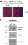
Smad-2 hyperactivation in Cav-1-null mammary epithelial cells. To assess the molecular mechanisms underlying the spontaneous EMT of Cav-1-deficient mammary epithelial cells, we evaluated the activation status of Smad-2. Cav-1 was previously shown to negatively regulate TGF-β signaling and to suppress Smad-2 phosphorylation in fibroblasts. After TGF-β stimulation, Smad-2 normally undergoes phosphorylation and translocates to the nucleus, resulting in gene activation. A: Immunofluorescence analysis with a monoclonal antibody raised against Smad-2 reveals that Smad-2 is localized to the nucleus in Cav-1-null mammary epithelial cells, suggesting that ablation of Cav-1 induces the constitutive activation of TGF-β signaling. B: WT and Cav-1 KO mammary epithelial cells were subjected to Western blot analysis with a phospho-specific probe directed against Smad-2. Note that Smad-2 is constitutively hyperphosphorylated in Cav-1-null cells. Equal loading was assessed by immunoblotting with a polyclonal antibody that recognizes total Smad-2. Immunoblotting with E-cadherin is shown as an additional control for equal epithelial protein content.
Figure 10
Figure 10
Cav-1-deficient acini exhibit increased branching in three-dimensional collagen cultures. To induce branching, single-cell suspensions of WT and Cav-1-null mammary epithelial cells were overlaid onto a mixture of Matrigel/collagen I (see Materials and Methods), where they formed acini. Starting from day 0, cells were either left untreated [no growth factor (GF) addition] or stimulated with HGF or with bFGF. It is important to note that EGF was omitted from the media. A: Quantitation of the percentage of branching epithelial structures. Note that in Cav-1 KO acini, HGF and bFGF stimulation induces an ∼1.7-fold and ∼1.9-fold increase in branching, respectively, as compared with WT controls. More than 70 acini were scored for each condition and for each genotype. *P ≤ 0.00003. Error bars, SEM. B–D: Phase images of WT and Cav-1-deficient acini grown in the absence of growth factors (B), in the presence of HGF (C), or in the presence of bFGF (D). Note that loss of Cav-1 increases the branching of these three-dimensional cultures of mammary epithelial cells. In C and D, three representative examples of the Cav-1 KO branching phenotype are shown. Arrows point at branching epithelial structures. Original magnifications, ×10.

References

    1. Hanahan D, Weinberg RA. The hallmarks of cancer. Cell. 2000;100:57–70. - PubMed
    1. Debnath J, Muthuswamy SK, Brugge JS. Morphogenesis and oncogenesis of MCF-10A mammary epithelial acini grown in three-dimensional basement membrane cultures. Methods. 2003;30:256–268. - PubMed
    1. Muthuswamy SK, Li D, Lelievre S, Bissell MJ, Brugge JS. ErbB2, but not ErbB1, reinitiates proliferation and induces luminal repopulation in epithelial acini. Nat Cell Biol. 2001;3:785–792. - PMC - PubMed
    1. Hay ED. An overview of epithelio-mesenchymal transformation. Acta Anat (Basel) 1995;154:8–20. - PubMed
    1. Santen RJ, Song RX, McPherson R, Kumar R, Adam L, Jeng MH, Yue W. The role of mitogen-activated protein (MAP) kinase in breast cancer. J Steroid Biochem Mol Biol. 2002;80:239–256. - PubMed

Publication types

MeSH terms

LinkOut - more resources