. 2006 Jan;5(1):18-25.
doi: 10.1128/EC.5.1.18-25.2006.

Developmentally regulated DNA methylation in Dictyostelium discoideum

Affiliations

Developmentally regulated DNA methylation in Dictyostelium discoideum

Mariko Katoh et al. Eukaryot Cell. 2006 Jan.

Abstract

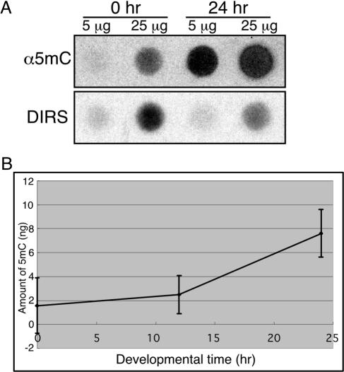
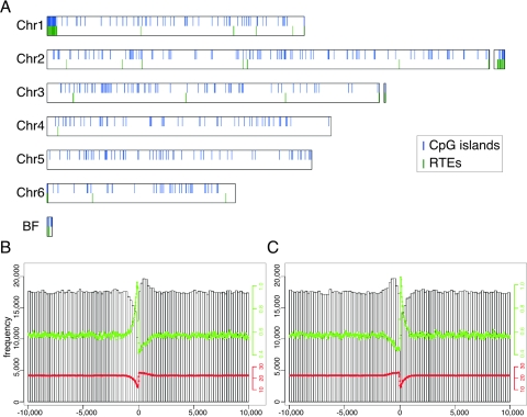
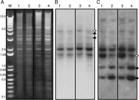
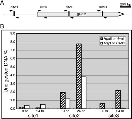
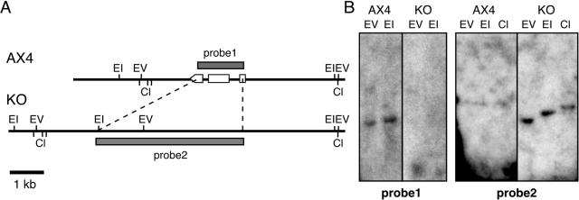
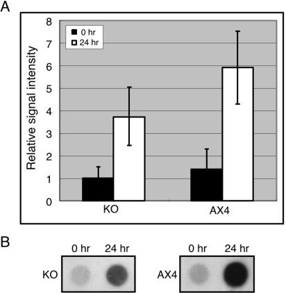
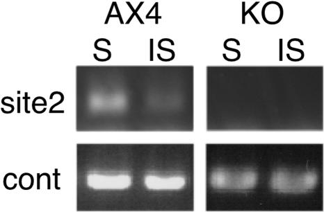
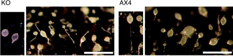
Methylation of cytosine residues in DNA plays a critical role in the silencing of gene expression, organization of chromatin structure, and cellular differentiation of eukaryotes. Previous studies failed to detect 5-methylcytosine in Dictyostelium genomic DNA, but the recent sequencing of the Dictyostelium genome revealed a candidate DNA methyltransferase gene (dnmA). The genome sequence also uncovered an unusual distribution of potential methylation sites, CpG islands, throughout the genome. DnmA belongs to the Dnmt2 subfamily and contains all the catalytic motifs necessary for cytosine methyltransferases. Dnmt2 activity is typically weak in Drosophila melanogaster, mouse, and human cells and the gene function in these systems is unknown. We have investigated the methylation status of Dictyostelium genomic DNA with antibodies raised against 5-methylcytosine and detected low levels of the modified nucleotide. We also found that DNA methylation increased during development. We searched the genome for potential methylation sites and found them in retrotransposable elements and in several other genes. Using Southern blot analysis with methylation-sensitive and -insensitive restriction endonucleases, we found that the DIRS retrotransposon and the guaB gene were indeed methylated. We then mutated the dnmA gene and found that DNA methylation was reduced to about 50% of the wild-type level. The mutant cells exhibited morphological defects in late development, indicating that DNA methylation has a regulatory role in Dictyostelium development. Our findings establish a role for a Dnmt2 methyltransferase in eukaryotic development.


Figures

FIG. 1.
FIG. 1.
DNA methylation is developmentally regulated. Genomic DNA was prepared from cells at different developmental stages and dot blotted on nitrocellulose membranes in the indicated amounts. (A) DNA methylation was detected by reacting the membranes with an antibody directed against 5-methylcytosine (α5mC), and the total amount of DNA was estimated by hybridization with a radioactive probe against DIRS-1 (DIRS). (B) The antibody and hybridization signals were quantified, and methylation levels were normalized to the amounts of DNA and plotted as a function of developmental time. The plot indicates that DNA methylation is increased during Dictyostelium development. Results are the averages and standard deviations from 3 replications.
FIG. 2.
FIG. 2.
Uneven distribution of CpG dinucleotides in the Dictyostelium genome. CpG islands were identified in the Dictyostelium genome. (A) CpG islands (blue vertical lines) and RTEs (green vertical lines) were plotted along the 6 chromosomes, indicating a high degree of overlap. All of the 13,629 predicted genes in the genome were aligned by their ORFs such that the translational start site (B) or the translational termination site (C) was aligned. CpG dinucleotide frequency was computed in a 20-kb region centered on the respective border of the ORF (open bars). The ratio between the observed and expected frequencies of CpG dinucleotides is shown as a green line. The G+C content (%) is indicated by a red line.
FIG. 3.
FIG. 3.
Selective DNA methylation in the DIRS retrotransposon and the guaB gene. Genomic DNA was prepared from vegetative (0 h, lanes 1 and 2) and developing (16 h, lanes 3 and 4) cells and digested with the XbaI restriction endonuclease and with restriction endonucleases that are sensitive (both HpaII and AvaI, lanes 1 and 3) or insensitive (both MspI and BsoBI, lanes 3 and 4) to 5mC. Digested DNA was separated by electrophoresis, along with the 1-kb DNA ladder as a size marker, and stained with ethidium bromide to visualize the banding pattern (A). The DNA was analyzed by Southern blotting with the DIRS probe (B) or with the guaB probe (C). Open arrows indicate the full-size fragments, and filled arrows indicate the resulting fragments.
FIG. 4.
FIG. 4.
Quantitative analysis of DNA methylation in the guaB locus. The guaB gene contains several putative methylation sites. (A) A physical map of the guaB locus with the 5′ border on the left. The ORF is indicated by a thick gray arrow. Quantitative PCR was done with four primer sets, indicated by small adjacent black arrows pointing at each other. Sites 1 and 3 are restriction sites for the methylation-sensitive endonuclease HpaII and the methylation-insensitive endonuclease MspI. Site 2 is a restriction site for the methylation-sensitive endonuclease AvaI and the methylation-insensitive endonuclease BsoBI. The control pair of primers (cont) was used for normalization. (B) Genomic DNA from vegetative (0 h) and terminally developed (24 h) cells was digested with methylation-sensitive (hatched bars) or -insensitive (white bars) endonucleases. The fraction (% of total) of DNA that remained undigested was measured by quantitative PCR across the respective sites as indicated in panel A and calculated from the average of two replications. Significant and developmentally regulated DNA methylation can be seen in sites 2 and 3.
FIG. 5.
FIG. 5.
Physical maps of the dnmA locus. (A) Restriction maps of the dnmA locus in the wild-type strain (AX4) and in the dnmA knockout strain (KO). Probe 1 consists of the dnmA gene, and probe 2 consists of the plasmid DNA used to replace the gene in the knockout mutant. Open boxes and arrowhead indicate the dnmA exons. Restriction sites are indicated as follows: EV, EcoRV; EI, EcoRI; Cl, ClaI. (B) Genomic DNA from the wild-type strain (AX4) and from the dnmA knockout strain (KO) was digested as indicated and subjected to Southern blot analysis with probes 1 and 2.
FIG. 6.
FIG. 6.
Reduced global DNA methylation in the dnmA knockout strain. Genomic DNA was extracted from vegetative (0 h) and developed (24 h) cells of the wild-type (AX4) and dnmA knockout (KO) strains. DNA methylation was detected by dot blotting with the anti-5mC antibody as described in the legend to Fig. 1. (A) DNA methylation signal intensity during growth (black bar) and development (white bar) are the averages of results from two independent replications of each of two independent knockout strains (KO) and the averages of results from three independent replications of the wild-type strain, showing a reduction in the developmentally regulated methylation. (B) Representative dot blot data comparing the dnmA knockout strain (KO) and the wild-type strain (AX4) signals in growth and development.
FIG. 7.
FIG. 7.
dnmA is responsible for selective DNA methylation at the guaB locus. Genomic DNA was prepared from developed wild-type (AX4) and dnmA knockout (KO) cells. The DNA was digested with a methylation sensitive (S, AvaI) or a methylation-insensitive (IS, BsoBI) endonuclease and analyzed by PCR using the primer set indicated in the legend to Fig. 4. PCR products were resolved on a 1% agarose gel containing ethidium bromide and visualized by fluorescence under UV light. Photographs are shown, and control PCR amplification products are shown below the experimental lanes.
FIG. 8.
FIG. 8.
dnmA is essential for proper development. Wild-type (AX4) and dnmA knockout (KO) cells were starved and developed on black nitrocellulose filters for 24 h. In the mutant cells, sori were found at various points along the stalk, whereas the wild type has one sorus at the top of the stalk. Mutant sori were also more transparent than wild-type sori.

References

    1. Adachi, H., T. Hasebe, K. Yoshinaga, T. Ohta, and K. Sutoh. 1994. Isolation of Dictyostelium discoideum cytokinesis mutants by restriction enzyme-mediated integration of the blasticidin S resistance marker. Biochem. Biophys. Res. Commun. 205:1808-1814. - PubMed
    1. Adams, R. L. 1996. DNA methylation, p. 33-66. In E. E. Bitter (ed.), Principles of medical biology, vol. 5. JAI Press, Inc., New York, N.Y.
    1. Aerts, S., G. Thijs, M. Dabrowski, Y. Moreau, and B. De Moor. 2004. Comprehensive analysis of the base composition around the transcription start site in Metazoa. BMC Genomics 5:34. - PMC - PubMed
    1. Bestor, T. H. 2000. The DNA methyltransferases of mammals. Hum. Mol. Genet. 9:2395-2402. - PubMed
    1. Bestor, T. H. 1998. The host defence function of genomic methylation patterns. Novartis Found. Symp. 214:187-199, 228-232. - PubMed

Publication types

LinkOut - more resources