. 2006 Jan;5(1):62-76.
doi: 10.1128/EC.5.1.62-76.2006.

Pair of unusual GCN5 histone acetyltransferases and ADA2 homologues in the protozoan parasite Toxoplasma gondii

Affiliations

Pair of unusual GCN5 histone acetyltransferases and ADA2 homologues in the protozoan parasite Toxoplasma gondii

Micah M Bhatti et al. Eukaryot Cell. 2006 Jan.

Abstract

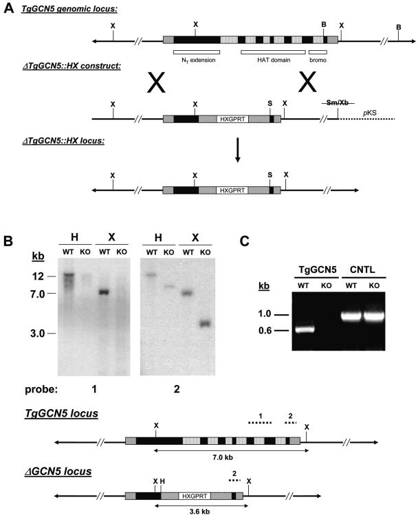
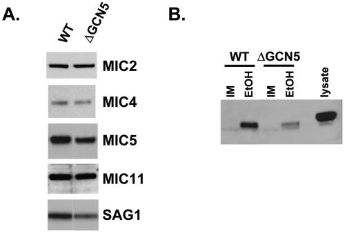
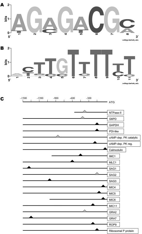
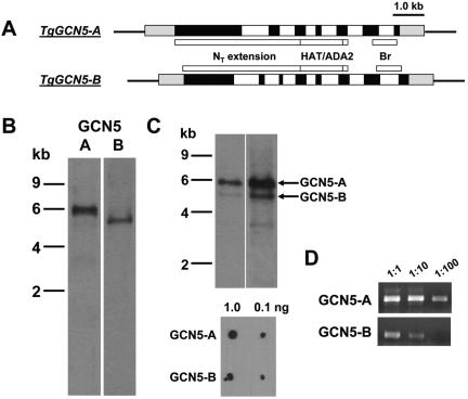
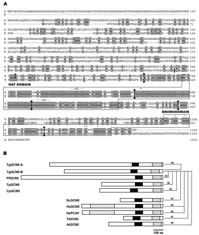
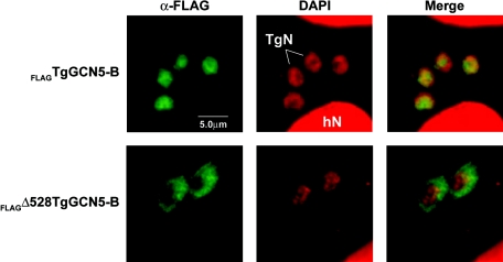
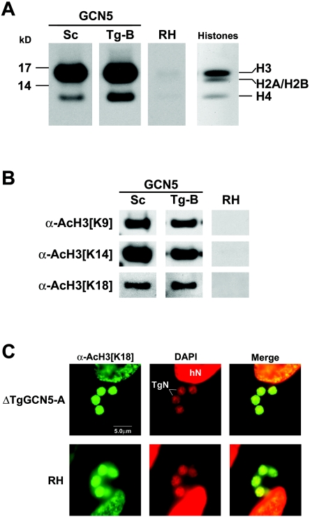
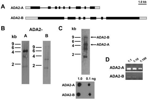
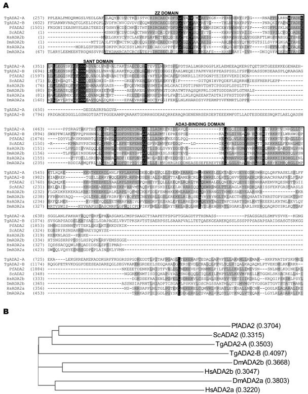
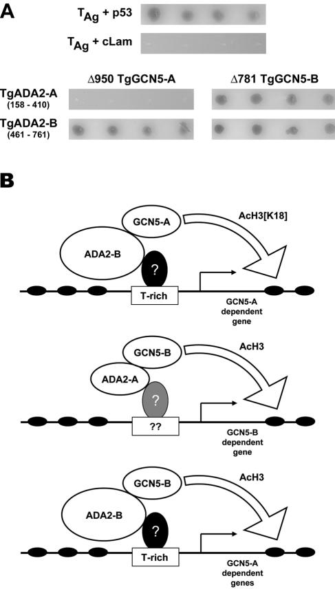
GCN5 is a histone acetyltransferase (HAT) essential for development in mammals and critical to stress responses in yeast. The protozoan parasite Toxoplasma gondii is a serious opportunistic pathogen. The study of epigenetics and gene expression in this ancient eukaryote has pharmacological relevance and may facilitate the understanding of these processes in higher eukaryotes. Here we show that the disruption of T. gondii GCN5 yields viable parasites, which were subsequently employed in a proteomics study to identify gene products affected by its loss. Promoter analysis of these TgGCN5-dependent genes, which were mostly parasite specific, reveals a conserved T-rich element. The loss of TgGCN5 does not attenuate virulence in an in vivo mouse model. We also discovered that T. gondii is the only invertebrate reported to date possessing a second GCN5 (TgGCN5-B). TgGCN5-B harbors a strikingly divergent N-terminal domain required for nuclear localization. Despite high homology between the HAT domains, the two TgGCN5s exhibit differing substrate specificities. In contrast to TgGCN5-A, which exclusively targets lysine 18 of H3, TgGCN5-B acetylates multiple lysines in the H3 tail. We also identify two ADA2 homologues that interact differently with the TgGCN5s. TgGCN5-B has the potential to compensate for TgGCN5-A, which probably arose from a gene duplication unique to T. gondii. Our work reveals an unexpected complexity in the GCN5 machinery of this primitive eukaryote.


Figures

FIG. 1.
FIG. 1.
Generation of ΔTgGCN5 parasite clone. (A) Schematic diagram comparing the endogenous genomic locus to the replacement allele contained on the ΔTgGCN5::HX “knockout” construct. Black boxes indicate exons, mottled boxes indicate introns, and gray boxes indicate UTRs. In a perfect allelic replacement via a double crossover event, virtually the entire TgGCN5 coding sequence would be replaced by the HXGPRT minigene cassette. Striped boxes denote the dihydrofolate reductase promoter and 3′ UTR regions. B, BglII; S, SpeI; Sm, SmaI; X, XhoI; Xb, XbaI. (B) Southern analysis verifies allelic replacement. Probe 1, a cDNA-derived portion of the HAT domain hybridizes to the expected size fragments in wild-type (WT) genomic DNA digested with indicated restriction enzymes. However, probe 1 does not hybridize to genomic DNA from the ΔTgGCN5 knockout clone (KO). Probe 2, designed to a portion of the final exon plus 3′ UTR, hybridizes to DNA fragments of the expected size in both WT and KO parasites. H, HindIII; X, XhoI. (C) TgGCN5 transcript is absent from ΔTgGCN5. Primers were designed to amplify a portion of the HAT domain of TgGCN5 using total RNA harvested from either wild-type (WT) or “knockout” (KO) parasites. Primers designed to amplify a control transcript (adenosine kinase) validate the RNA purification from each sample. CNTL, control.
FIG. 2.
FIG. 2.
(A) Western analysis comparing levels of indicated protein between wild-type (WT) and ΔTgGCN5 parasites. Samples were normalized to protein concentration. Antibody dilutions applied were as follows (dilutions are indicated in parentheses): anti-SAG1 (1:500), anti-MIC2 (1:10,000), anti-MIC4 (1:5,000), anti-MIC5 (1:5,000), anti-MIC11 (1:1,000). (B) Immunoblot for MIC2 following a microneme secretion assay. IM, invasion media (control); EtOH, ethanol, used at a final concentration of 1%. Parasite lysate displays the size of unprocessed MIC2 for comparison.
FIG. 3.
FIG. 3.
Promoter analysis of candidate GCN5-dependent genes. (A) A known cis-acting enhancer of T. gondii genes (33) was identified by MEME in some of the promoters of genes reportedly down-regulated in ΔTgGCN5. The letter size depicts the relative abundance of each nucleotide conserved in the sequence. (B) T-rich element present in GCN5-dependent promoters analyzed in Table 1. (C) Schematic diagram of promoter sequences showing approximate location of the T-rich element. All positions are indicated with respect to the translational start site. A triangle indicates the position of the motif; gray triangles signify that the motif was found on the opposite strand. PDI, protein disulfide isomerase; cAMP, cyclic AMP; PK, protein kinase; dep, dependent; reg, regulatory.
FIG. 4.
FIG. 4.
A pair of GCN5 HATs in T. gondii. (A) Schematic diagram of the genomic loci for TgGCN5-A and -B. TgGCN5-A is comprised of 7 exons (black) and 6 introns (white), while TgGCN5-B contains 8 exons. The approximate location of key domains in the encoded protein are indicated. HAT, histone acetyltransferase domain; ADA2, ADA2-binding domain; Br, bromodomain. Gray boxes represent untranslated regions. (B) Northern blots probed for TgGCN5-A and -B transcripts, respectively. (C) Dual-probing of a single Northern blot with both TgGCN5s. Two different exposure times are shown. Lower panel shows that equivalent amounts of nonisotopically labeled probes emit approximately equal chemiluminescent signals, so the differences observed on the blot are not due to a bias in probe labeling. (D) Equal amounts of cDNA generated from wild-type RNA were used in PCRs to amplify either TgGCN5-A or -B.
FIG. 5.
FIG. 5.
TgGCN5-B protein analysis. (A) Pairwise alignment of the deduced protein sequences for TgGCN5-A and -B. Dark gray highlights show identical residues; light gray denotes conserved differences. Locations of introns in the genomic sequence corresponding to the protein are indicated by vertical lines with diamondheads at junction sites. HAT and bromodomains are boxed and labeled. The conserved glutamic acid residue critical for HAT activity is marked with an asterisk, and conserved bromodomain residues important for interacting with acetylated lysines are designated with plus signs. (B) Cartoon depicting protein structures of GCN5 family proteins from Apicomplexa (top 5) and other eukaryotes (lower 5). Black boxes indicate HAT domains, gray boxes indicate bromodomains, and mottled boxes indicate PCAF homology domains. ADA2-binding domains are located between HAT and bromodomains. Numerical values represent the percent identities between TgGCN5-B and the indicated protein. Tg, Toxoplasma gondii; Pf, Plasmodium falciparum; Tp, Theileria parvum; Cp, Cryptosporidium parvum; Sc, Saccharomyces cerevisiae; Hs, Homo sapiens; Tt, Tetrahymena thermophila; At, Arabidopsis thaliana. It should be noted that structures for C. parvum and T. parvum were drawn based on predicted open reading frames in genome sequencing databases.
FIG. 6.
FIG. 6.
TgGCN5-B localizes to the parasite nucleus via the N-terminal extension. Recombinant protein in parasites expressing either full-length FLAGTgGCN5-B or FLAGΔ528TgGCN5-B was detected by immunofluorescence assay. Antibody recognizing the FLAG epitope is shown in green. Infected cells were also stained with 4′,6′-diamidino-2-phenylindole (DAPI), shown in red. hN, host cell nucleus; TgN, parasite nucleus.
FIG. 7.
FIG. 7.
TgGCN5-B preferentially acetylates histone H3 at multiple lysine residues. (A) Autoradiogram of HAT assays using core histones and either recombinant yeast GCN5 (Saccharomyces cerevisiae, Sc) or FLAGTgGCN5-B (Tg-B). An equivalent amount of untransfected parasite lysate was used as a negative control. The right panel is a Coomassie-stained SDS-polyacrylamide gel showing the positions of the core histones. (B) Western blot of similar HAT assays performed on recombinant histone H3, probed with antibodies (α) to specific acetylated (Ac) H3 lysine residues. (C) Immunofluorescence assay showing acetylation of H3 [K18] in ΔTgGCN5 and wild-type (RH) parasites (green). 4′,6′-Diamidino-2-phenylindole (DAPI) was applied to stain nuclei (red). hN, host cell nucleus; TgN, parasite nucleus.
FIG. 8.
FIG. 8.
A pair of ADA2s in T. gondii. (A) Schematic diagram of the genomic loci for TgADA2-A and -B. Black boxes represent exons, and the lines in between are introns. Gray boxes represent untranslated regions. (B) Northern blots probed for TgADA2-A and -B transcripts, ∼4 kb and ∼10 kb, respectively. Bands at ∼1.7 and 3.5 kb overlap with rRNA contamination. (C) Dual probing of a single Northern blot with both TgADA2s. The lower panel shows that equivalent amounts of nonisotopically labeled probes emit approximately equal chemiluminescent signals. (D) Equal amounts of cDNA generated from wild-type RNA were used in PCRs to amplify either TgADA2-A or -B.
FIG. 9.
FIG. 9.
Comparative analysis of ADA2 domains. (A) ADA2 domains from TgADA2-A and -B are aligned for comparison to those found in other species. Black boxes denote identity of residues across species; gray boxes denote residues that are chemically similar. The ZZ, SANT, and ADA3-binding domains are boxed. Numerals in parentheses refer to amino acid numbers. Tg, Toxoplasma gondii; Pf, Plasmodium falciparum; Sc, Saccharomyces cerevisiae; Hs, Homo sapiens; Dm, Drosophila melanogaster. (B) Phylogenic analysis of ADA2 orthologues from representative species with the calculated distance values in parentheses.
FIG. 10.
FIG. 10.
Protein-protein interactions between TgGCN5s and TgADA2s. (A) Top panels: AH109 yeast were cotransformed with plasmids expressing simian virus 40 large-T antigen (TAg) and murine p53 (positive control) or TAg and human lamin C (cLam) (negative control). Four independent colonies initially plated on TDO plates were spotted onto QDO plates containing X-α-gal and grown for 3 days at 30°C. Growth of blue colonies represents protein-protein interaction. Bottom panels: AH109 yeast were cotransformed with the plasmids indicated and plated for incubation as described above. All colonies shown are blue in color. (B) Working model for TgGCN5 HAT complexes. Based on yeast two-hybrid data, at least three possible complexes containing GCN5 may exist in T. gondii. The lowest panel depicts a complex comprised of TgGCN5-B and TgADA2-B that could compensate for TgGCN5-A. Based on what is observed in other eukaryotes, a question mark refers to a putative DNA-binding transcription factor with the capacity to recruit the GCN5 complexes by virtue of binding ADA2. Note that the position of the T-rich motif is approximate, as its actual location varies as shown in Fig. 3C.

Similar articles

Cited by

References

    1. Bailey, T. L., and C. Elkan. 1994. Fitting a mixture model by expectation maximization to discover motifs in biopolymers. Proc. Int. Conf. Intell. Syst. Mol. Biol. 2:28-36. - PubMed
    1. Barlev, N. A., R. Candau, L. Wang, P. Darpino, N. Silverman, and S. L. Berger. 1995. Characterization of physical interactions of the putative transcriptional adaptor, ADA2, with acidic activation domains and TATA-binding protein. J. Biol. Chem. 270:19337-19344. - PubMed
    1. Berger, S. L., B. Pina, N. Silverman, G. A. Marcus, J. Agapite, J. L. Regier, S. J. Triezenberg, and L. Guarente. 1992. Genetic isolation of ADA2: a potential transcriptional adaptor required for function of certain acidic activation domains. Cell 70:251-265. - PubMed
    1. Bhatti, M. M., and W. J. Sullivan, Jr. 2005. Histone acetylase GCN5 enters the nucleus via importin-alpha in protozoan parasite Toxoplasma gondii. J. Biol. Chem. 280:5902-5908. - PubMed
    1. Black, M. W., and J. C. Boothroyd. 2000. Lytic cycle of Toxoplasma gondii. Microbiol. Mol. Biol Rev. 64:607-623. - PMC - PubMed

Publication types

MeSH terms

Associated data

LinkOut - more resources