. 2006 Jan;5(1):192-202.
doi: 10.1128/EC.5.1.192-202.2006.

SST2, a regulator of G-protein signaling for the Candida albicans mating response pathway

Affiliations

SST2, a regulator of G-protein signaling for the Candida albicans mating response pathway

Daniel Dignard et al. Eukaryot Cell. 2006 Jan.

Abstract

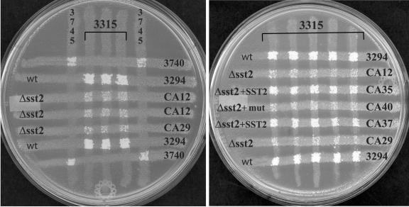
Candida albicans contains a functional mating response pathway that is similar to the well-studied system of Saccharomyces cerevisiae. We have characterized a regulator of G protein signaling (RGS) homolog in C. albicans with sequence similarity to the SST2 gene of Saccharomyces cerevisiae. Disruption of this gene, which had been designated SST2, causes an opaque MTLa/MTLa derivative of strain SC5314 to show hypersensitivity to the C. albicans alpha-factor. This hypersensitivity generates an enhanced cell cycle arrest detected in halo assays but reduces the overall mating efficiency of the cells. Transcriptional profiling of the pheromone-regulated gene expression in the sst2 mutant shows a pattern of gene induction similar to that observed in wild-type cells, but the responsiveness is heightened. This involvement of an RGS in the sensitivity to pheromone is consistent with the prediction that the mating response pathway in C. albicans requires the activation of a heterotrimeric G protein.


Figures

FIG. 1.
FIG. 1.
Disruption of SST2. (A) PCR-based cassette method for disruption of SST2 in 2 steps. The thick black bar represents genomic DNA at the SST2 locus, and the white rectangles represent the SST2 gene coding sequence. The PCR cassettes used for the disruption are composed of a selectable marker (gray ovoid rectangle) flanked by two 80-nucleotide segments from the SST2 locus (small white rectangles) for the homologous recombination of the cassettes. The first allele of SST2 was replaced by the HIS1 marker, and the second allele was replaced by the URA3 marker. Small arrows represent orientation and approximate position of oligonucleotides (Table 2) used for PCR analysis and confirmation of the disruption. (B) Confirmation of disruption by PCR. The parent strain 3294 (lanes 1 to 8), the first allele disrupted strain CA3 (lanes 9 to 16), and the second allele disrupted strain CA12 (lanes 17 to 24) were analyzed by PCR and amplified with the oligonucleotides identified in white over the upper part of the agarose gel (PCR). A 1-kb DNA ladder (Invitrogen, Carlsbad, CA) was used for size reference (lanes M). PCR with oligonucleotides 1 and 2 produce a 2.44-kb DNA fragment for the SST2 wild-type allele, as seen for strains 3294 and CA3 (lanes 1 and 9) but absent in Δsst2 strain CA12 (lane 17), a 1.76-kb fragment when SST2 is replaced by the HIS1 marker, as seen for strains CA3 and CA12 (lanes 9 and 17), or a 1.86-kb fragment in strain CA12 when the second SST2 allele is replaced by the URA3 marker (lane 17). The 1.76-kb and the 1.86-kb PCR fragments from the two markers were not well resolved on this gel (lane 17), so the presence of the two markers in this PCR product was confirmed by digestion with specific restriction enzymes, as described in Materials and Methods. The deletion of the SST2 gene was also confirmed by PCR with SST2 internal oligonucleotides 3 and 4. No PCR band is visible for Δsst2 strain CA12 (lane18), confirming that no other copy of the SST2 gene is detectable, while the 1.1-kb internal fragment is visible for strains 3294 and CA3 (lanes 2 and 10). The proper integration sites of the two markers are demonstrated in the other lanes.
FIG. 2.
FIG. 2.
DNA microarray data for genes induced by α-factor in Δsst2 strain. Each of the 11 conditions consists of an experiment (EXP) compared to a reference (ref) whose description is summarized above the column of values. The description includes the strain name (full genotypes are given in Table 1; CA12 is designated Δsst2), the strain mating type (MTL) as either MTLa (a) or MTLα (alpha), and the strain phase as either opaque (Op) or white (wh). In conditions 3 to 7, cultures were induced by pheromone, either the synthetic α-factor peptide 13α (+13) (conditions 4 to 7) or, in condition 3, by factors in the supernatant from the opposite mating type culture: supernatant from MTLa culture (+“a”) or supernatant from MTLα culture (+“α”). Genes induced more than twofold by α-factor in the Δsst2 strain are shown for the 11 conditions as signal intensity EXP/ref ratios in black boxes if the ratio is >2.0 and in gray-shaded boxes if the ratio is <0.5. Genes are identified by their orf19 number and annotation names. Controls were added for an opaque-phase-specific gene ops4, and opaque MTLα-specific gene MFalpha1. The data were clustered for conditions having similar transcription profiles as depicted by the dendrogram above the table.
FIG. 3.
FIG. 3.
RGS genes in C. albicans. Analysis of the C. albicans genome revealed 2 ORFs with an RGS domain. Schematic representation of the 2 ORFs, orf19.695 and orf19.4222, aligned with the most similar gene from S. cerevisiae. Rectangles represent RGS domains, and ovoid rectangles represent DEP domains, identified using SMART (32). The first DEP domain of ScSST2 is part of a more generalized fungal DR domain (31). The length of each ORF, based on the predicted amino acid sequence, is shown at the right of each arrow.
FIG. 4.
FIG. 4.
Halo assay. Improved growth arrest of Δsst2 strains in the presence of mating pheromone α-factor. For the assay, SC plates were seeded with 1 × 10⁵ cells from a colony, and 5 μl of α-factor peptide 13α at 1 μg/μl was spotted over the black dot (13) or 5 μl of solvent for the negative control (control). (A) Wild-type parent strain 3294; (B) Δsst2 strain CA12; (C) Δsst2 strain CA12 in the white phase; (D) Δsst2 strain CA29; (E) strain CA37, Δsst2 plus wild-type SST2; (F) strain CA40, Δsst2 plus mutant sst2. Cells were in the opaque phase, except for those in panel C. The wild-type parent strain 3294 produces weak halos only (A). The halos are significantly better after deletion of the SST2 gene (B and D), and reintegration of a wild-type copy of the SST2 gene reverts this phenotype (E). Reintegration of a mutant sst2 gene (see Materials and Methods) does not suppress the phenotype of Δsst2 strains (F).
FIG. 5.
FIG. 5.
Reduced mating of Δsst2 strains. Strains of opposite mating types were crossed for 24 h on YPD plates for mating by auxotrophic marker complementation (see Materials and Methods), and cells were transferred by replica to plates lacking 5 amino acids for selection of the mating products. The selective plates are shown after 2 days incubation. The petri plates are labeled with each strain used in the assays and are described in Table 1. Reduced mating, relative to wild-type parent strain 3294, is observed for strains CA12, CA29, and CA40, and near wild-type mating efficiency is observed for the SST2-reintegrated strains CA35 and CA37. Strains 3740 and 3745 were included as controls; they have, respectively, the same auxotrophic markers as strains 3294 and 3315 but are of opposite mating types.
FIG. 6.
FIG. 6.
Pheromone-induced morphological changes. The α-factor peptide 13α was added at different concentrations to liquid culture for the wild-type parent strain (3294) and for the Δsst2 strain (CA12). The concentration of α-factor in the cultures at t0 is indicated at the left; the reference, consisting of the addition of solvent without peptide, is marked 0 μg/ml. Typical unconstricted projections (shmoos) are highlighted with white arrows. Many cells develop projections in the presence of α-factor at 0.1 μg/ml, and the two strains are undistinguishable. However, morphological change is difficult to detect for strain 3294 at a lower concentration of α-factor (0.01 μg/ml), while Δsst2 cells are still responsive at 0.001 μg/ml. Pictures are shown at a magnification of ×1,000, 6 h after a single-dose addition of peptide to cells grown in SC medium.

Similar articles

Cited by

References

    1. Achstetter, T. 1989. Regulation of alpha-factor production in Saccharomyces cerevisiae: a-factor pheromone-induced expression of the MFα1 and STE13 genes. Mol. Cell. Biol. 9:4507-4514. - PMC - PubMed
    1. Bennett, R. J., M. A. Uhl, M. G. Miller, and A. D. Johnson. 2003. Identification and characterization of a Candida albicans mating pheromone. Mol. Cell. Biol. 23:8189-8201. - PMC - PubMed
    1. Bernards, A., and J. Settleman. 2004. GAP control: regulating the regulators of small GTPases. Trends Cell Biol. 14:377-385. - PubMed
    1. Cabrera-Vera, T. M., J. Vanhauwe, T. O. Thomas, M. Medkova, A. Preininger, M. R. Mazzoni, and H. E. Hamm. 2003. Insights into G protein structure, function, and regulation. Endocr. Rev. 24:765-781. - PubMed
    1. Chan, R. K., and C. A. Otte. 1982. Isolation and genetic analysis of Saccharomyces cerevisiae mutants supersensitive to G1 arrest by a factor and α factor pheromones. Mol. Cell. Biol. 2:11-20. - PMC - PubMed

Publication types

LinkOut - more resources