. 2006 Jan 17;45(2):523-32.
doi: 10.1021/bi051235w.

Role of Tyr348 in Tyr385 radical dynamics and cyclooxygenase inhibitor interactions in prostaglandin H synthase-2

Affiliations

Role of Tyr348 in Tyr385 radical dynamics and cyclooxygenase inhibitor interactions in prostaglandin H synthase-2

Corina E Rogge et al. Biochemistry. .

Abstract

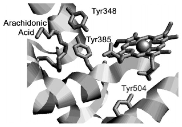
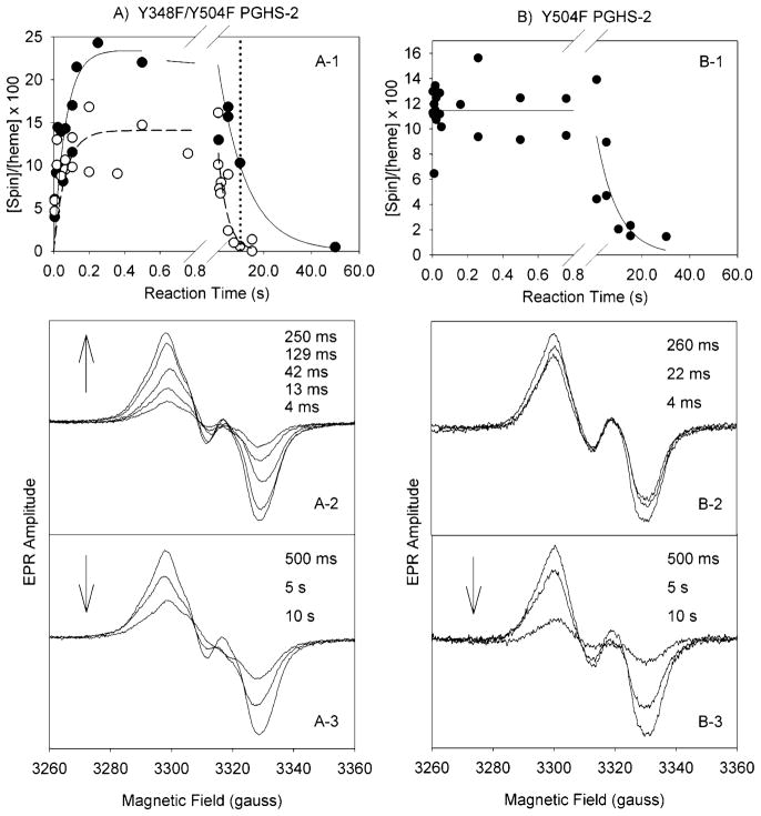
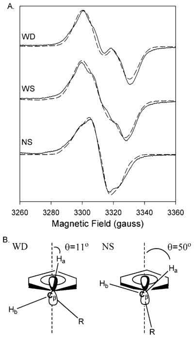
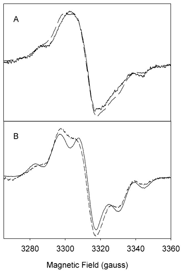
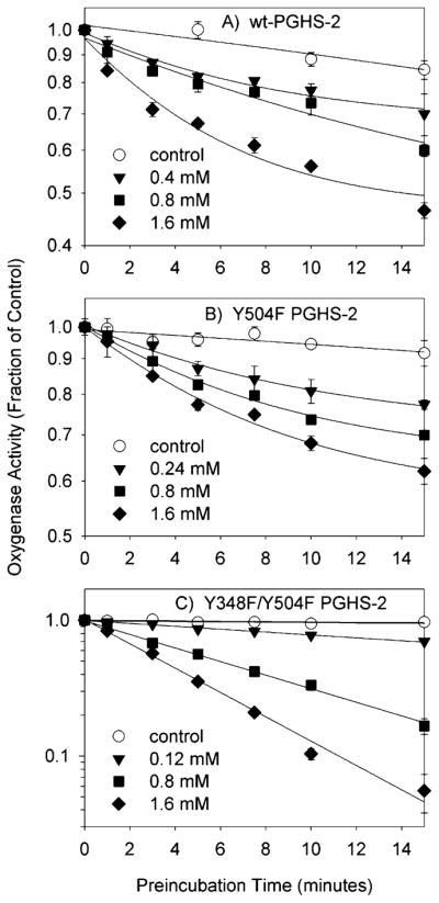
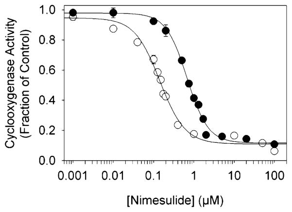
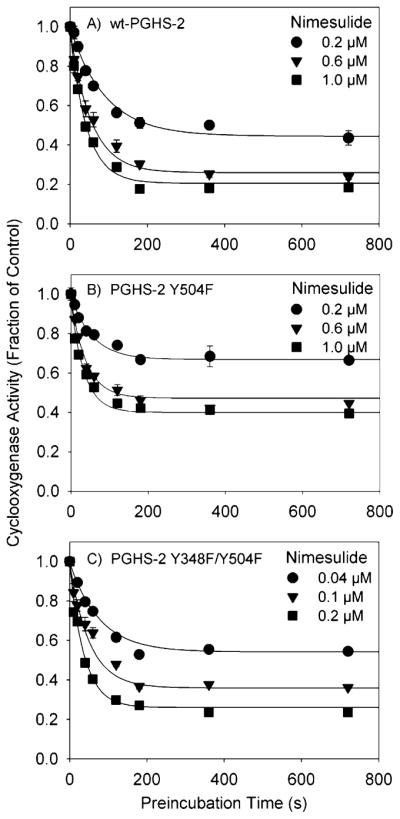
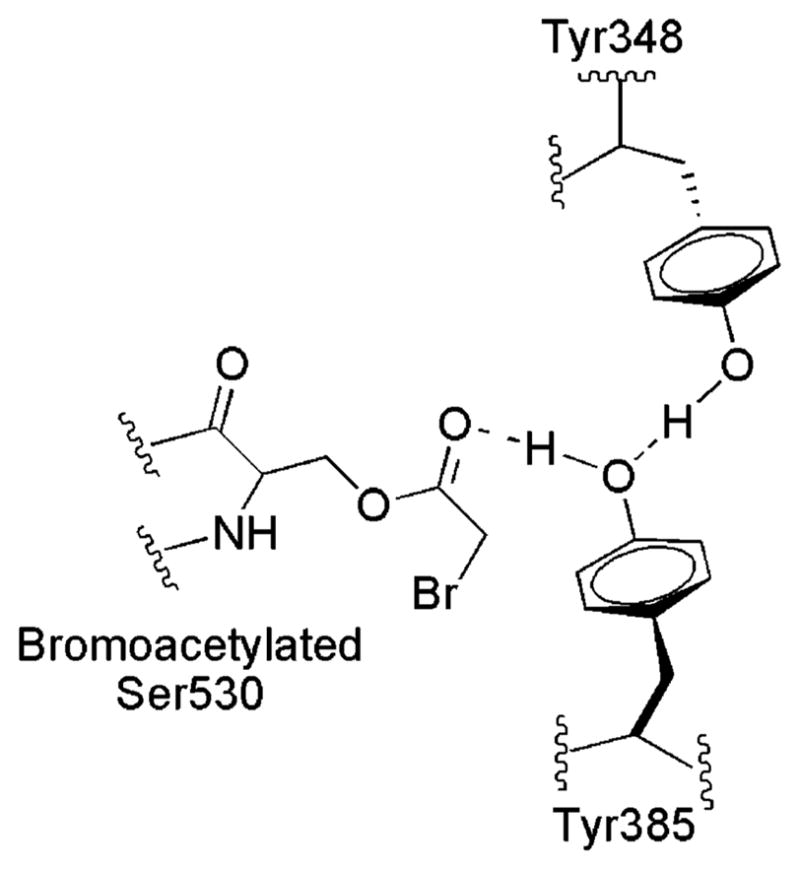
Both prostaglandin H synthase (PGHS) isoforms utilize a radical at Tyr385 to abstract a hydrogen atom from arachidonic acid, initializing prostaglandin synthesis. A Tyr348-Tyr385 hydrogen bond appears to be conserved in both isoforms; this hydrogen bonding has the potential to modulate the positioning and reactivity of the Tyr385 side chain. The EPR signal from the Tyr385 radical undergoes a time-dependent transition from a wide doublet to a wide singlet species in both isoforms. In PGHS-2, this transition results from radical migration from Tyr385 to Tyr504. Localization of the radical to Tyr385 in the recombinant human PGHS-2 Y504F mutant was exploited in examining the effects of blocking Tyr385 hydrogen bonding by introduction of a further Y348F mutation. Cyclooxygenase and peroxidase activities were found to be maintained in the Y348F/Y504F mutant, but the Tyr385 radical was formed more slowly and had greater rotational freedom, as evidenced by observation of a transition from an initial wide doublet species to a narrow singlet species, a transition not seen in the parent Y504F mutant. The effect of disrupting Tyr385 hydrogen bonding on the cyclooxygenase active site structure was probed by examination of cyclooxygenase inhibitor kinetics. Aspirin treatment eliminated all oxygenase activity in the Y348F/Y504F double mutant, with no indication of the lipoxygenase activity observed in aspirin-treated wild-type PGHS-2. Introduction of the Y348F mutation also strengthened the time-dependent inhibitory action of nimesulide. These results suggest that removal of Tyr348-Tyr385 hydrogen bonding in PGHS-2 allows greater conformational flexibility in the cyclooxygenase active site, resulting in altered interactions with inhibitors and altered Tyr385 radical behavior.


Figures

Figure 1
Figure 1
Crystal structure of oPGHS-1 complexed with arachidonic acid (PDB entry 1DIY), showing the relative positions of Tyr385, Tyr348, Tyr504, arachidonic acid, and CoPPIX (14).
Figure 2
Figure 2
Tyrosyl radical kinetics during the reaction of Y348F/Y504F PGHS-2 and Y504F PGHS-2 with EtOOH. (A) The double mutant (68 μM heme) in 100 mM KPi (pH 7.2), 50 μM phenol, 0.04% octyl glucoside, and 10% glycerol was reacted at room temperature with 15 equiv of EtOOH. (A-1) Time course of Y348F/Y504F tyrosyl radical intensity as determined by double integration of the EPR signals (●) in experiments with two separate enzyme preparations and time course for the PGHS-2 Y148F/Y348F/Y404F/Y504F quadruple mutant (○) (25). (A-2 and A-3) EPR spectra from the double mutant reaction samples freeze trapped at the indicated times. (B) Y504F PGHS-2 (48 μM heme) was reacted with 15 equiv of EtOOH at room temperature (25). (B-1) Time course of radical intensity determined by double integration. (B-2 and B-3) EPR spectra from the Y504F mutant reaction samples freeze trapped at the indicated times.
Figure 3
Figure 3
(A) Experimental (—) and simulated (– – –) EPR spectra of the doublet, wide singlet (WS), and narrow singlet (NS) EPR signals from Y348F/Y504F PGHS-2 reacted with peroxide. The doublet was observed after reaction of the double mutant (38 μM heme) with 15 equiv of EtOOH for 104 ms. The simulated spectrum represents the sum of 80% of the Y504F WD simulated spectrum and 20% of the NS simulated spectrum. The WS was observed after reaction of the double mutant (15 μM heme) with 15 equiv of EtOOH on ice for ~10 s. The wide singlet simulation represents the sum of 48% simulated Y504F WD and 52% simulated NS. The NS spectrum was recorded after preincubation of the double mutant (15 μM heme) with 5 equiv of nimesulide and subsequent reaction with 15 equiv of EtOOH for 10 s on ice. Parameters for simulation of the Y504F WD and NS spectra are given in Table 3. (B) Structural diagrams of the proposed conformations of the β-methylene groups of tyrosyl radicals that give rise to the WD and NS spectra.
Figure 4
Figure 4
EPR spectra of radicals formed during anaerobic reaction of Y348F/Y504F PGHS-2 with AA or d8-AA. (A) Reaction at 0 °C of Y348F/Y504F PGHS-2 (15 μM heme) with 15 equiv of EtOOH for 26 s and then with 4 equiv of AA for 11 s (—). The simulated EPR spectrum (– – –) is an arithmetic combination of 59% Y348F/Y504F PGHS-2 NS and 41% AA-pentadienyl radical. (B) Reaction at 0 °C of Y348F/Y504F PGHS-2 (15 μM heme) with 15 equiv of EtOOH for 19 s and then with 4 equiv of d8-AA for 13 s (—). The simulated EPR spectrum (– – –) is an arithmetic combination of 54% Y348F/Y504F PGHS-2 NS and 46% d8-AA-pentadienyl radical. Simulation parameters are given in Table 3 and ref .
Figure 5
Figure 5
Kinetics of cyclooxygenase inhibition by aspirin in wild-type PGHS-2 and the Y504F and Y348F/Y504F mutants. The initial velocity of oxygen consumption was measured after preincubation of PGHS-2 (A), the Y504F mutant (B), or the Y348F/Y504F mutant (C) with the indicated levels of aspirin for the indicated times. Data shown are the averages of triplicate measurements, normalized to the uninhibited control; standard deviations are indicated by error bars. Lines represent exponential or linear fits to the data.
Figure 6
Figure 6
Cyclooxygenase inhibition by nimesulide in PGHS-2 (●) and the Y348F/Y504F double mutant (○). The enzyme was preincubated for 1 min at 30 °C with the indicated level of nimesulide before the reaction was initiated by addition of arachidonate (100 μM). Activities are normalized to uninhibited control values; data represent averages of triplicate measurements, and standard deviations are indicated by error bars.
Figure 7
Figure 7
Kinetics of cyclooxygenase inhibition by nimesulide in PGHS-2 and the Y504F and Y348F/Y504F mutants. The initial velocity of oxygen consumption was measured after preincubation of PGHS-2 (A), the Y504F mutant (B), or the Y348F/Y504F double mutant (C) with the indicated levels of nimesulide for the indicated times. Data are the averages of triplicate measurements, normalized to the control; standard deviations are given by error bars. The curves represent theoretical fits to the data using the model described by Callan et al. (30).
Figure 8
Figure 8
EPR spectra of tyrosyl radicals formed during reaction of PGHS-2, Y348F/Y504F PGHS-2, and PGHS-1 with EtOOH. Individual EPR spectra are scaled to the same amplitude and overlaid to facilitate line shape comparisons. (A) PGHS-2 (11.2 μM heme) in 100 mM KPi (pH 7.2), 0.1% Tween-20, and 10% glycerol was reacted at room temperature with 5 equiv of EtOOH for the indicated times. Data are from ref . (B) The Y348F/Y504F PGHS-2 double mutant (68 μM heme) in 100 mM KPi (pH 7.2), 50 μM phenol, 0.04% octyl glucoside, and 10% glycerol was reacted at room temperature with 15 equiv of EtOOH for the indicated times. (C) PGHS-1 (8.5 μM heme) in 100 mM KPi (pH 7.2), 0.1% Tween-20, and 10% glycerol was reacted at room temperature with 5 equiv of EtOOH for the indicated times. Data are from ref .
Figure 9
Figure 9
Schematic structure of bromo-acetylated oPGHS-1 (PDB entry 1PTH) depicting the interactions among Tyr385, Tyr348, Ser530, and the bromoacetyl group based on crystallographic data from ref .

Similar articles

Cited by

References

    1. Smith WL, Dewitt DL. Prostaglandin endoperoxide H synthases-1 and -2. Adv Immunol. 1996;62:167–215. - PubMed
    1. Herschman HR. Prostaglandin synthase 2. Biochim Biophys Acta. 1996;1299:125–140. - PubMed
    1. Lambeir AM, Markey CM, Dunford HB, Marnett LJ. Spectral properties of the higher oxidation states of prostaglandin H synthase. J Biol Chem. 1985;260:14894–14896. - PubMed
    1. Tang MS, Copeland RA, Penning TM. Detection of an Fe2+-protoporphyrin-IX intermediate during aspirin-treated prostaglandin H2 synthase II catalysis of arachidonic acid to 15-HETE. Biochemistry. 1997;36:7527–7534. - PubMed
    1. Lu G, Tsai AL, Van Wart HE, Kulmacz RJ. Comparison of the peroxidase reaction kinetics of prostaglandin H synthase-1 and -2. J Biol Chem. 1999;274:16162–16167. - PubMed

Publication types

MeSH terms

LinkOut - more resources