CytoregR inhibits growth and proliferation of human adenocarcinoma cells via induction of apoptosis
- PMID: 16401338
- PMCID: PMC1343545
- DOI: 10.1186/1477-3163-5-1
CytoregR inhibits growth and proliferation of human adenocarcinoma cells via induction of apoptosis
Abstract
Background: Cancer is one of the devastating neovascular diseases that incapacitate so many people the world over. Recent reports from the National Cancer Institute indicate some significant gain therapy and cancer management as seen in the increase in the 5-year survival rate over the past two decades. Although near-perfect cure rate have been reported in the early-stage disease, these data reveal high recurrence rate and serious side effects including second malignancies and fatalities. Most of the currently used anticancer agents are only effective against proliferating cancer cells. Thus attention has been focused on potential anti-cancer agents capable of killing cancer cells independent of the cell cycle state, to ensure effective elimination of most cancer cells. The objective of this study was to test the chemosensitivity and potential mechanism of action of a novel cancer drug, CytoregR, in a panel of human cancer cells.
Methods: the study was performed using a series of bioassays including Trypan blue exclusion, MTS Growth inhibition, LDH-cytotoxicity, TUNEL-Terminal DNA fragmentation Apoptosis Assay, and the Caspase protease CPP32 activity assays.
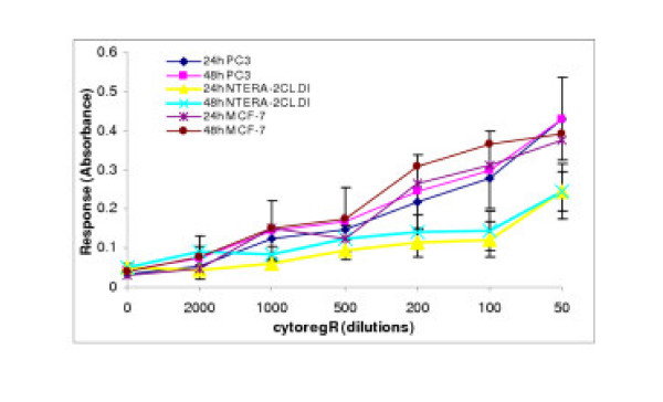
Results: CytoregR induced significant dose- and time-dependent inhibition of growth in all the cells; with significant differences in chemosensitivity (P < 0.05) between the target cells becoming more apparent at 48 hr exposure. CytoregR showed no significant effect on normal cells relative to the tumor cells. Growth inhibition in all the cells was due to induction of apoptosis at lower concentrations of cytoregR (> 1:300). CytoregR-induced caspase protease-3 (CPP32) activation significantly and positively correlated with apoptosis induction and growth inhibition; thus implicating CPP32 as the principal death pathway in cytoregR-induced apoptosis.
Conclusion: CytoregR exerted a dose-and time-dependent growth inhibitory effect in all the target cells through induction of apoptosis via the CPP32 death pathway, independent of hormonal sensitivity of the cells. The present data indicate that not only could CPP32 provide a potential target for regulation of cytoregR-induced apoptosis but also that cytoregR could play a significant role in chemotherapeutic regimen in many human malignant tumors.
Figures


Similar articles
-
Wild-type p53-dependent etoposide-induced apoptosis mediated by caspase-3 activation in human glioma cells.J Neurosurg. 2000 Aug;93(2):289-97. doi: 10.3171/jns.2000.93.2.0289. J Neurosurg. 2000. PMID: 10930016
-
A novel hydroxamic acid compound, BMD188, demonstrates anti-prostate cancer effects by inducing apoptosis. I: In vitro studies.Anticancer Res. 1999 Jan-Feb;19(1A):51-60. Anticancer Res. 1999. PMID: 10226524
-
Immunohistochemistry of Caspase3/CPP32 in human stomach and its correlation with cell proliferation and apoptosis.Anticancer Res. 1998 Nov-Dec;18(6A):4347-53. Anticancer Res. 1998. PMID: 9891491
-
TNF-alpha induces apoptosis mediated by AEBSF-sensitive serine protease(s) that may involve upstream caspase-3/CPP32 protease activation in a human gastric cancer cell line.Int J Oncol. 2000 Jun;16(6):1243-8. Int J Oncol. 2000. PMID: 10812002
-
Potential mechanism of phytochemical-induced apoptosis in human prostate adenocarcinoma cells: Therapeutic synergy in genistein and beta-lapachone combination treatment.Cancer Cell Int. 2004 Aug 17;4(1):5. doi: 10.1186/1475-2867-4-5. Cancer Cell Int. 2004. PMID: 15315711 Free PMC article.
Cited by
-
lncRNA CASC9 positively regulates CHK1 to promote breast cancer cell proliferation and survival through sponging the miR‑195/497 cluster.Int J Oncol. 2019 May;54(5):1665-1675. doi: 10.3892/ijo.2019.4734. Epub 2019 Feb 28. Int J Oncol. 2019. PMID: 30816435 Free PMC article.
-
Effect of an ionic antineoplastic agent Cytoreg on blood chemistry in a Wistar rat model.Med Gas Res. 2022 Jan-Mar;12(1):18-23. doi: 10.4103/2045-9912.324592. Med Gas Res. 2022. PMID: 34472498 Free PMC article.
-
Unveiling the physicochemical, photocatalytic, antibacterial and antioxidant properties of MWCNT-modified Ag2O/CuO/ZnO nanocomposites.RSC Adv. 2025 Jan 16;15(2):1323-1334. doi: 10.1039/d4ra08466g. eCollection 2025 Jan 9. RSC Adv. 2025. PMID: 39822572 Free PMC article.
-
Evaluation of dermal corrosion and irritation by Cytoreg in rabbits.Toxicol Rep. 2021 Jul 31;8:1527-1529. doi: 10.1016/j.toxrep.2021.07.021. eCollection 2021. Toxicol Rep. 2021. PMID: 34408971 Free PMC article.
References
-
- Jemal A, Clegg LX, Ward E, Ries LAG, Wu X, Jamison PM, Wingo P, Howe HL, Anderson RN, Edwards BK. Annual Report to the Nation on the Status of Cancer, 1975–2001, with a Special Feature Regarding Survival. Cancer. 2004 http://www.cdc.gov/cancer, http://www.cancer.gov/newscenter/pressreleases/ReportNation2004Release, http://interscience.wiley.com/cancer/report2004 - PubMed
-
- American Cancer Society Report to the nation on the status of cancer. Retrieved from http://www.cancer.org/eprise/main/docroot/STT/stt 0,2003
-
- Shottenfeld D, Warshauer M, Sherlock S, Zauher A, Leder M, Payne R. The epidermiology of testicular cancer in young adults. Am J Epidemiol. 1980;112:232–246. - PubMed
-
- Brown GG. Testicular cancer: an overview. Med Surg Nursing. 2003;12:37–48. - PubMed
LinkOut - more resources
Full Text Sources
Research Materials
Miscellaneous