. 2006 Jan 10:5:2.
doi: 10.1186/1477-3163-5-2.

Expression of hyaluronan in human tumor progression

Affiliations

Expression of hyaluronan in human tumor progression

Rajeev K Boregowda et al. J Carcinog. .

Abstract

Background: The development and progression of human tumors is accompanied by various cellular, biochemical and genetic alterations. These events include tumor cells interaction with extracellular matrix molecules including hyaluronan (HA). Hyaluronan is a large polysaccharide associated with pericellular matrix of proliferating, migrating cells. Its implication in malignant transformation, tumor progression and with the degree of differentiation in various invasive tumors has well accepted. It has been well known the role HA receptors in tumor growth and metastasis in various cancer tissues. Previously we have observed the unified over expression of Hyaluronic Acid Binding Protein (HABP), H11B2C2 antigen by the tumor cells in various types progressing tumor tissues with different grades. However, the poor understanding of relation between HA and HA-binding protein expression on tumor cells during tumor progression as well as the asymmetric observations of the role of HA expression in tumor progression prompted us to examine the degree of HA expression on tumor cells vs. stroma in various types of human tumors with different grades.

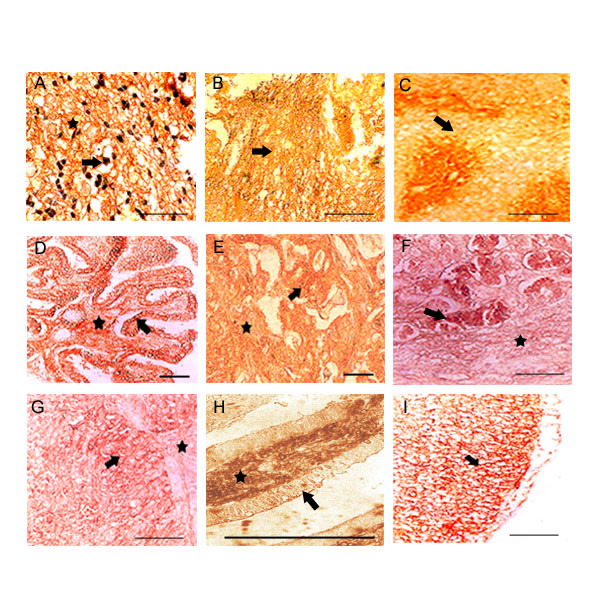
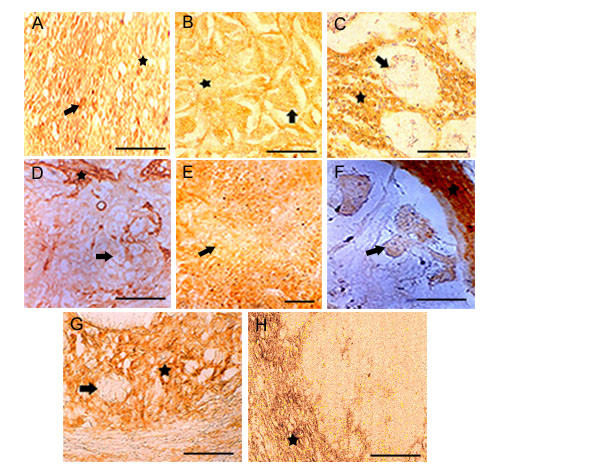
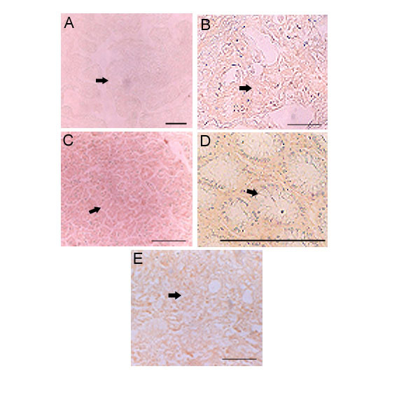
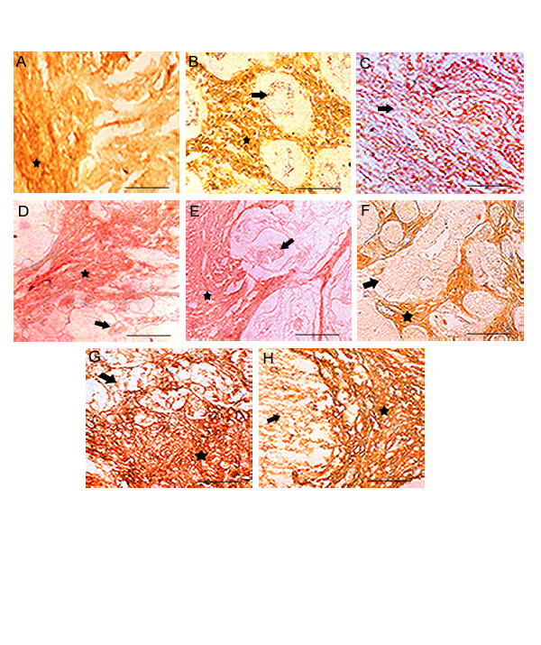
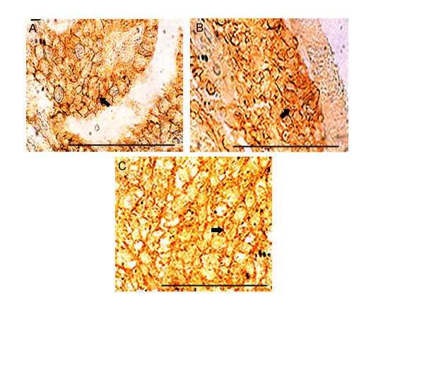
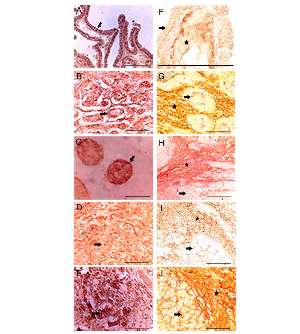
Methods: In the present study clinically diagnosed tumor tissue samples of different grades were used to screen the histopathological expression of hyaluronan by using b-PG (biotinylated proteoglycan) as a probe and we compared the relative HA expression on tumor cells vs. stroma in well differentiated and poorly differentiated tumors. Specificity of the reaction was confirmed either by pre-digesting the tissue sections with hyaluronidase enzyme or by staining the sections with pre-absorbed complex of the probe and HA-oligomers.

Results: We show here the down regulation of HA expression in tumor cells is associated with progression of tumor from well differentiated through poorly differentiated stage, despite the constant HA expression in the tumor associated stroma.

Conclusion: The present finding enlighten the relative roles of HA expression on tumor vs. stroma during the progression of tumors.


Figures

Figure 1
Figure 1
HA expression in well differentiated tumors: Only 8 examples are shown. Top row, (A, B, C) represents cerebral astrocytomas, ganglioglioma and salivary gland tumor. In astrocytomas neuroglial fibers and the astrocyte membrane are highly reactive for HA. Neoplastic nerve cells with extensive fibrillar processes are HA positive in ganglioglioma. Benign epithelia surrounding the tumor cells in salivary of showed low HA reaction. Middle row, (D, E, F) represents papillary carcinoma of thyroid, infiltrating breast and stomach tumor. Thyroid tumor showed intense HA staining at intraepithelial and in surrounding stromal components. High level HA expression was observed in tumor cells and surrounding stroma in case of infiltrating breast tumor. Tumor epithelia of stomach showed strong HA reaction. Bottom row, (G, H, I) represents Transitional epithelia of urinary bladder, descending colon and 5 days old chicken embryo limb. Transitional epithelia of urinary bladder showed intense staining at intra-epithelial and in surrounding tumor components. Invasive colon tumor cells showed strong HA reaction. 5 days chicken embryo limb was used as positive control, where the mesoderm is intensely positive for HA. Arrow shows the tumor cells and the asterisk shows stroma. Scale bars, 50 μm.
Figure 2
Figure 2
HA expression in poorly differentiated tumors: Only 8 examples are shown. Top row, (A, B, C) shows disappearance of HA stain from the tumor epithelia. Fibrillar astrocytomas show intense reaction while astrocytes are negative. Infiltrating breast and stomach tumor epithelia shows the loss of HA while the stromal components shows intense HA stain. Middle row, (D, E F) shows the loss HA expression in urinary bladder, pancreas (tumor epithelia) and caecum tumor epithelia while the stromal areas from urinary bladder and caecum were enriched with HA positive materials. Bottom row, (G, H) represents prostate and ovary. Prostate is showing numerous invading tumor cells in stromal areas. Only the stromal components are stained with b.PG. Ovary shows total absence of HA expression in tumor cells while the stroma is enriched with HA. Arrow shows the tumor cells and the asterisk shows stroma. Scale bars, 50 μm.
Figure 3
Figure 3
Specificities of b.PG for HA staining: Only 5 examples are shown. A. Thyroid, B. Infiltrating breast, C. Pancreas, D. Colon and E. Urinary bladder. All tumors are negative for Hyaluronic acid under different control experimental condition (explained in the material and methods). Scale bars, 50 μm.
Figure 4
Figure 4
HA staining in poorly differentiated stromal areas: Top row, (A, B, C) represents breast, stomach and gallbladder. Middle row, (D, E, F) represents colon, caecum and prostate. Bottom row, (G, H) represents urinary bladder and renal cell carcinoma. The stromal and intratumoral areas are highly positive for HA in all tumors, where as the loss of HA expression observed in the tumor cells. Arrow shows the tumor cells and the asterisk shows stroma. Scale bars, 50 μm.
Figure 5
Figure 5
HA on tumor epithelial cell surfaces: Three examples are shown. (A, B, C). A. Thyroid, B. Descending Colon, C. Urinary bladder. Tumors derived from well differentiated stage. Only 3 examples are shown. High accumulation of HA reactive materials observed on the cell surfaces of all the tumors. Arrows shows the cell surface. Scale bars, 50 μm.
Figure 6
Figure 6
Comparison of HABP and HA expression in poorly differentiated tumors: HABP expression in poorly differentiated tumors (A, B, C, D, E), A. Thyroid, B. Stomach, C. Descending Colon, D. Urinary bladder, E. Pancreas shows high HABP expression both on tumor cell surface and nucleus. HA expression in poorly differentiated tumors (F, G, H, I, J), F. Thyroid, G. Stomach, H. Descending Colon, I. Urinary bladder, J. Pancreas shows very low HA expression in tumor cells where as the tumor stroma shows high expression of HA. Arrow shows the tumor cells and the asterisk shows stroma. Scale bars, 50 μm.

References

    1. Toole BP. Hyaluronan: From extracellular glue to pericellular cue. Nature Rev Cancer. 2004;4:528–539. doi: 10.1038/nrc1391. - DOI - PubMed
    1. Yoneda M. Key molecules to an understanding of intracellular hyaluronan function. Conn Tissue. 2001;33:227–233.
    1. Evanko SP, Wight TN. Intracellular localization of hyaluronan in proliferating cells. J Histochem Cytochem. 1999;47:1331–1342. - PubMed
    1. Toole BP. In: Hyaluronan, in proteoglycans; structure, biology and molecular interactions. Iozzo RV, editor. Marcel Dekker, New York; 2000. pp. 61–92.
    1. Grammatikakis N, Grammatikakis A, Yoneda M, Yu Q, Banerjee SD, Toole BP. A novel glycosaminoglycan-binding protein is the vertebrate homologue of the cell cycle control protein Cdc37. J Biol Chem. 1995;270:16198–16205. doi: 10.1074/jbc.270.27.16198. - DOI - PubMed