. 2006 Jan;130(1):104-19.
doi: 10.1053/j.gastro.2005.10.012.

Sustained phosphorylation of Bid is a marker for resistance to Fas-induced apoptosis during chronic liver diseases

Affiliations

Sustained phosphorylation of Bid is a marker for resistance to Fas-induced apoptosis during chronic liver diseases

Arndt Vogel et al. Gastroenterology. 2006 Jan.

Abstract

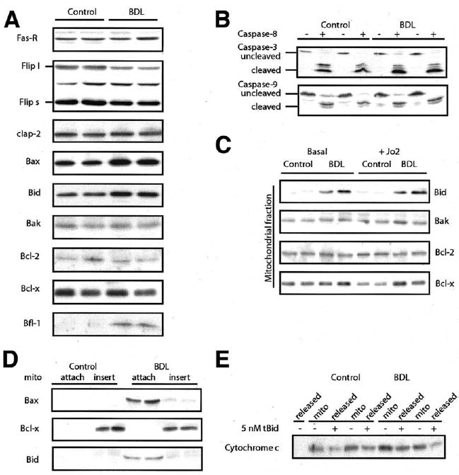
Background & aims: Increased rates of apoptosis have been reported to play a role in the pathophysiology of many disorders, including liver diseases. Conversely, genetic mutations that result in impairment of programmed cell death have been associated with cancer development. However, apoptosis resistance can also be the result of nongenetic stress adaptation, as seen in the cancer-prone metabolic liver disease hereditary tyrosinemia. To clarify whether stress-induced apoptosis resistance is a general feature of chronic liver diseases, an animal model of chronic cholestasis was examined.

Methods: Studies were performed with mice before and 2 weeks following bile duct ligation and with Fah-/- and Fah/p21-/- mice before and after NTBC withdrawal.

Results: Here we show that bile duct ligation induced profound resistance against Fas monoclonal antibody-mediated hepatocyte death. The apoptosis signaling pathway was blocked downstream of caspase-8 activation and proximal to mitochondrial cytochrome c release. In controls, activation of the Fas receptor resulted in rapid dephosphorylation of Bid and its subsequent cleavage, whereas Bid remained phosphorylated and uncleaved in chronic cholestasis and other models of hepatic apoptosis resistance.

Conclusions: We propose a model in which the phosphorylation status of Bid determines the apoptotic threshold of hepatocytes in vivo. Furthermore, resistance to apoptosis in chronic cholestasis may contribute to the long-term risk of cancer in this setting.


Figures

Figure 1
Figure 1
Bile duct–ligated mice develop a resistance against Fas-induced apoptosis. (A) Wild-type mice underwent BDL and were challenged with the Fas mAb Jo2 (0.35 μg/g) 14 days later. Survival of control (squares) and bile duct–ligated (triangles) mice are shown. All control mice (n = 10) died from acute liver failure within 12 hours following injection of Jo2, whereas all bile duct–ligated mice survived (n = 10). (B) DNA laddering following challenge with Jo2 was only seen in control mice. Results are shown for duplicate animals under each condition. A standard DNA ladder marker (m) is shown. (C) TUNEL staining (brown color) of liver sections is shown (original magnification 200×) in control and bile duct–ligated mice before and after Jo2 injection. TUNEL-positive cells were only visible in control mice following Jo2 injection.
Figure 2
Figure 2
The mitochondrial pathway is blocked in bile duct–ligated mice. (A) Liver injury after Jo2 challenge in control (WT; open columns) (n = 4) and bile duct–ligated mice (filled columns) (n = 4). Alanine aminotransferase and bilirubin levels were measured in plasma before (F.0) and after (F.4) challenge with Jo2. Alanine aminotransferase levels increased only in control mice following Jo2 injection. Bilirubin levels were significantly elevated in bile duct–ligated mice. Caspase-8 and caspase-3 activities were determined in liver homogenates 2 hours (F.2; caspase 8) or 4 hours (F.4; caspase 3) following Jo2 injection (n = 4 each). In both groups, activation of caspase-8 was observed, whereas caspase-3 activity was only detectable in control mice. (B) Western blots of Bid, Bax, cytochrome c, and caspase-9 before (basal) and 4 hours after (Fas) induction of apoptosis with Jo2 in control and bile duct–ligated mice (n = 4 each; 2 of 4 samples shown here) to determine activation of the mitochondrial pathway of apoptosis. Cleavage of Bid (decrease of full-length Bid), translocation of Bax, release of cytochrome c, and cleavage of caspase-9 following Jo2 occurred only in control mice. F.0, before Fas mAb injection, F.2/F.4, 2/4 hours following Fas mAb injection.
Figure 3
Figure 3
Analysis of the mitochondrial pathway in wild-type and bile duct–ligated mice. (A) Western blot analysis of liver tissues in control and bile duct–ligated mice in the basal state without Fas mAb injection. Shown are data from 2 of 4 mice in each group. Liver tissues were analyzed for total cellular levels of Fas-R, Flips/l, cIap-2, Bax, Bid, Bak, Bcl-2, Bcl-x, and A1/Bfl-1. Of all the proteins analyzed, only A1/Bfl-1 was significantly induced 2 weeks following BDL. (B) Liver extracts of control and bile duct–ligated mice were incubated with in vitro activated recombinant caspase-8. Western blots for in vitro cleavage of caspase-3 and caspase-9 revealed no difference in both groups. (C) The mitochondrial fraction of control and bile duct–ligated mice was analyzed for BH-3–like proteins. Levels of Bak, Bcl-2, and Bcl-x were not significantly changed before and after challenge with Jo2, whereas Bid was significantly increased in this fraction 2 weeks following BDL even before Jo2 injection. (D) Alkaline extraction of mitochondria from bile duct–ligated mice and controls. Bid and Bax were only loosely attached and not inserted into the mitochondrial membrane of bile duct–ligated mice in contrast to Bcl-x. (E) Isolated liver mitochondria (n = 4 each; 2 of 4 samples shown here) were incubated with different concentrations of in vitro cleaved tBid, and translocation of cytochrome c from the mitochondrial (mito) fraction to the cytosol (released) was monitored by immunoblotting with a cytochrome c antibody. Cytochrome c release following incubation with 5 nmol/L tBid is shown. First row (−), spontaneous cytochrome c release without tBid incubation; second to ninth row, the overall mitochondrial cytochrome c content of the respective sample (n = 2 per group) is shown followed by the released cytosolic cytochrome c fraction following incubation with tBid. There was no difference in cytochrome c release between bile duct–ligated mice and controls.
Figure 4
Figure 4
Bid is not dephosphorylated following Fas injection in bile duct–ligated and in Fah−/−/p21−/− mice. (A) Phosphorylation of Bid at S61 was measured in cytosolic liver extracts from control and bile duct–ligated mice by Western blot. Increased phosphorylation of Bid in bile duct–ligated mice (n = 4 each; 2 of 4 samples shown here) before challenge with Jo2 and, more importantly, no dephosphorylation following Jo2 injection were seen. In contrast, Bid was almost completely dephosphorylated following induction of apoptosis in control mice. (B) Western blot analysis of CK1ε and PP2A levels in cell extracts from control and bile duct–ligated mice. The cytosolic CK1ε levels were higher in bile duct–ligated mice. Levels of PP2A were not changed in cytosolic extracts (data not shown). (C) CK1 activity was measured in cell extracts of control and bile duct–ligated mice with a CK1-specific peptide (n = 4). Note that activity assay is not specific for CK1ε but measures overall CK1 activity. CK1 activity was higher in bile duct–ligated mice at all time points. Interestingly, CK1 activity was reduced in control mice following challenge with Jo2, but this reduction was not seen in bile duct–ligated mice. (D) Cellular extracts of control and bile duct–ligated mice following Jo2 injection were analyzed for in vitro phosphorylation activity of His6-Bid (representative blot, n = 4 each); negative control (−) contains no cell extract, and positive control (CK1) contains recombinant CK1. Shown is a Western blot of the membranes probed with the phosphor-specific Bid (S61) antibody. Phosphorylation of Bid was only detectable in bile duct–ligated mice and was almost absent in controls. CK1 inhibitor (CKI-7) significantly inhibited phosphorylation of His6-Bid in bile duct–ligated mice. Total His6-Bid levels of non—CKI-7–treated reactions are shown. (E) Fah−/− and Fah−/−/p21−/− mice were challenged with Jo2 2 weeks following NTBC withdrawal. Dephosphorylation and subsequent cleavage of Bid and cytochrome c release occurred only in the double-knockout mice. Total CK1ε levels were unchanged, whereas cytosolic levels were lower in Fah−/−/p21−/− mice following Jo2 injection. (F) Wild-type mice were injected with Jo2 alone or concomitantly with suramin. Suramin prevents dephosphorylation of Bid and subsequent Bid cleavage and cytochrome c release. Cytosolic CK1ε levels are higher in suramin-treated mice. (G) In suramin-treated mice, only a few scattered TUNEL-positive hepatocytes were visible following Jo2 injection.
Figure 5
Figure 5
Atm phosphorylates Bid in vitro but is not necessary to prevent Fas-induced apoptosis during chronic liver diseases. (A) The ATM-p53 DNA damage pathway is activated in bile duct–ligated mice, measured by increased phosphorylation of p53 and increased overall levels of p53 and effector proteins p21 and Bax. (B) Recombinant Atm phosphorylated Bid-GST in vitro. (C) Bile duct–ligated Atm−/− mice are protected against Fas mAb–induced apoptosis similar to wild-type mice. Dephosphorylation of Bid and subsequent cleavage of Bid, release of cytochrome c, and cleavage of caspase-9 and caspase-3 occurs only in wild-type mice. (D and E) TUNEL staining (brown color) of liver sections is shown (original magnification 200×). TUNEL-positive cells are only visible (D) in control mice 6 hours after Jo2 injection but not (E) in Atm−/− mice. (F) Cytosolic CK1ε levels are again higher in bile duct–ligated mice, whereas PP2A levels remain unchanged.

Similar articles

Cited by

  • Principles of therapeutic liver repopulation.
    Grompe M. Grompe M. J Inherit Metab Dis. 2006 Apr-Jun;29(2-3):421-5. doi: 10.1007/s10545-006-0311-2. J Inherit Metab Dis. 2006. PMID: 16763912 Review.
  • Protection from rapamycin-induced apoptosis by insulin-like growth factor-I is partially dependent on protein kinase C signaling.
    Thimmaiah KN, Easton JB, Houghton PJ. Thimmaiah KN, et al. Cancer Res. 2010 Mar 1;70(5):2000-9. doi: 10.1158/0008-5472.CAN-09-3693. Epub 2010 Feb 23. Cancer Res. 2010. PMID: 20179209 Free PMC article.
  • Apoptotic cell death in disease-Current understanding of the NCCD 2023.
    Vitale I, Pietrocola F, Guilbaud E, Aaronson SA, Abrams JM, Adam D, Agostini M, Agostinis P, Alnemri ES, Altucci L, Amelio I, Andrews DW, Aqeilan RI, Arama E, Baehrecke EH, Balachandran S, Bano D, Barlev NA, Bartek J, Bazan NG, Becker C, Bernassola F, Bertrand MJM, Bianchi ME, Blagosklonny MV, Blander JM, Blandino G, Blomgren K, Borner C, Bortner CD, Bove P, Boya P, Brenner C, Broz P, Brunner T, Damgaard RB, Calin GA, Campanella M, Candi E, Carbone M, Carmona-Gutierrez D, Cecconi F, Chan FK, Chen GQ, Chen Q, Chen YH, Cheng EH, Chipuk JE, Cidlowski JA, Ciechanover A, Ciliberto G, Conrad M, Cubillos-Ruiz JR, Czabotar PE, D'Angiolella V, Daugaard M, Dawson TM, Dawson VL, De Maria R, De Strooper B, Debatin KM, Deberardinis RJ, Degterev A, Del Sal G, Deshmukh M, Di Virgilio F, Diederich M, Dixon SJ, Dynlacht BD, El-Deiry WS, Elrod JW, Engeland K, Fimia GM, Galassi C, Ganini C, Garcia-Saez AJ, Garg AD, Garrido C, Gavathiotis E, Gerlic M, Ghosh S, Green DR, Greene LA, Gronemeyer H, Häcker G, Hajnóczky G, Hardwick JM, Haupt Y, He S, Heery DM, Hengartner MO, Hetz C, Hildeman DA, Ichijo H, Inoue S, Jäättelä M, Janic A, Joseph B, Jost PJ, Kanneganti TD, Karin M, Kashkar H, Kaufmann T, Kelly … See abstract for full author list ➔ Vitale I, et al. Cell Death Differ. 2023 May;30(5):1097-1154. doi: 10.1038/s41418-023-01153-w. Epub 2023 Apr 26. Cell Death Differ. 2023. PMID: 37100955 Free PMC article. Review.
  • Targeting cancer cells by ROS-mediated mechanisms: a radical therapeutic approach?
    Trachootham D, Alexandre J, Huang P. Trachootham D, et al. Nat Rev Drug Discov. 2009 Jul;8(7):579-91. doi: 10.1038/nrd2803. Epub 2009 May 29. Nat Rev Drug Discov. 2009. PMID: 19478820 Review.
  • At the crossroads of homoeostasis and disease: roles of the PACS proteins in membrane traffic and apoptosis.
    Youker RT, Shinde U, Day R, Thomas G. Youker RT, et al. Biochem J. 2009 Jun 12;421(1):1-15. doi: 10.1042/BJ20081016. Biochem J. 2009. PMID: 19505291 Free PMC article. Review.

References

    1. Galle PR, Krammer PH. CD95-induced apoptosis in human liver disease. Semin Liver Dis. 1998;18:141–151. - PubMed
    1. Vogel A, van Den Berg IE, Al-Dhalimy M, Groopman J, Ou CN, Ryabinina O, Iordanov MS, Finegold M, Grompe M. Chronic liver disease in murine hereditary tyrosinemia type 1 induces resistance to cell death. Hepatology. 2004;39:433–443. - PubMed
    1. Findor J, He XS, Sord J, Terg R, Gershwin ME. Primary biliary cirrhosis and hepatocellular carcinoma. Autoimmun Rev. 2002;1:220–225. - PubMed
    1. Caballeria L, Pares A, Castells A, Gines A, Bru C, Rodes J. Hepatocellular carcinoma in primary biliary cirrhosis: similar incidence to that in hepatitis C virus-related cirrhosis. Am J Gastroenterol. 2001;96:1160–1163. - PubMed
    1. Keeffe EB, Pinson CW, Ragsdale J, Zonana J. Hepatocellular carcinoma in arteriohepatic dysplasia. Am J Gastroenterol. 1993;88:1446–1449. - PubMed

Publication types