. 2006 Jan 16;172(2):309-20.
doi: 10.1083/jcb.200507067. Epub 2006 Jan 9.

Receptor palmitoylation and ubiquitination regulate anthrax toxin endocytosis

Affiliations

Receptor palmitoylation and ubiquitination regulate anthrax toxin endocytosis

Laurence Abrami et al. J Cell Biol. .

Abstract

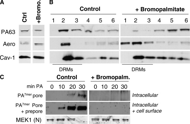
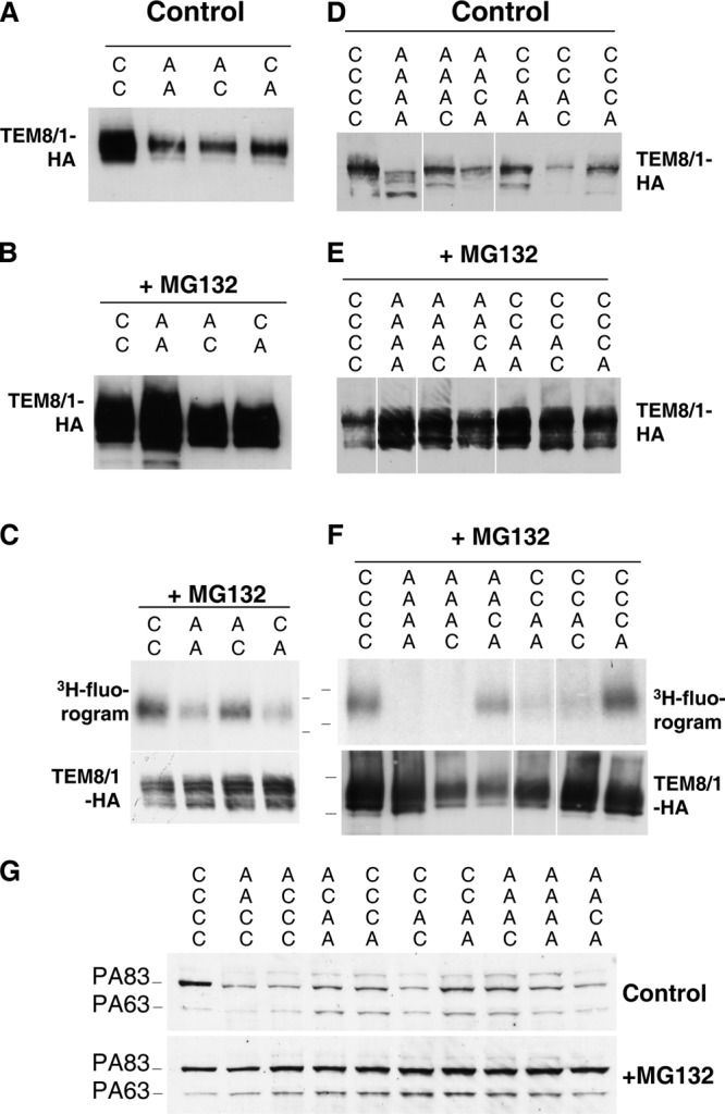
The anthrax toxin is composed of three independent polypeptide chains. Successful intoxication only occurs when heptamerization of the receptor-binding polypeptide, the protective antigen (PA), allows binding of the two enzymatic subunits before endocytosis. We show that this tailored behavior is caused by two counteracting posttranslational modifications in the cytoplasmic tail of PA receptors. The receptor is palmitoylated, and this unexpectedly prevents its association with lipid rafts and, thus, its premature ubiquitination. This second modification, which is mediated by the E3 ubiquitin ligase Cbl, only occurs in rafts and is required for rapid endocytosis of the receptor. As a consequence, cells expressing palmitoylation-defective mutant receptors are less sensitive to anthrax toxin because of a lower number of surface receptors as well as premature internalization of PA without a requirement for heptamerization.


Figures

Figure 1.
Figure 1.
TEM8/1 and CMG2/4 are glycosylated. (A) Alignment of the cytoplasmic tails of human TEM8 and human CMG2 using the SIM software of the EXPASY server (www.expasy.ch). Regions of identity are shown in yellow, lysine residues in green, and cysteine residues in red. Numbering of the residues corresponds to that of TEM8/1. The asterisks label the lysine mutants in the K6R mutant described in Fig. 7. (B and C) CHOΔATR cells untransfected and stably or transiently transfected (for 48 h; B) with a TEM8/1-HA construct were analyzed by Western blotting (40 μg of protein/lane; B) or immunofluorescence (C) using an anti-HA antibody. Bar, 10 μM. (D) CHOΔATR cells stably expressing TEM8/1-HA were submitted or unsubmitted to surface trypsinization at 4°C and subsequently analyzed for the presence of TEM8/1-HA by Western blotting. (E) CHOΔATR cells transiently expressing TEM8/1-HA were grown in the presence or absence of tunicamycin. Control cell extracts were subsequently left untreated or treated with N-glycosidase F or Endo H. The effects of these treatments were analyzed by SDS-PAGE followed by Western blotting against the HA tag. (B and E) Band u, unglycosilated form; p, glycosylated precursor; m, mature form. (F) Extracts of CHOΔATR cells transiently expressing or not expressing CMG2/4-V5 were left untreated or treated with N-glycosidase F or Endo H and subsequently analyzed by SDS-PAGE followed by Western blotting against the V5 tag.
Figure 2.
Figure 2.
Palmitoylation events are required for DRM association and internalization of PA. (A and B) Control BHK cells were pretreated or untreated with bromopalmitate and were incubated with 500 ng/ml nicked PA63 and 20 ng/ml aerolysin for 1 h at 4°C followed by 10 min at 37°C. (A) Cell extracts were submitted to SDS-PAGE followed by Western blotting to reveal PA63, aerolysin, and caveolin-1 (Cav-1). (B) Cells were solubilized in 1% Triton X-100, run on an OptiPrep gradient, and each fraction was analyzed by SDS-PAGE followed by Western blotting against PA, aerolysin, and caveolin-1. (C) BHK cells were pretreated with bromopalmitate and incubated with 500 ng/ml nicked PA63 for 1 h at 4°C followed by different times at 37°C. Cell extracts (40 μg of protein) were analyzed by SDS-PAGE and Western blotting to reveal the SDS-resistant PA7mer pore and MEK1 (NH2-terminal directed). To detect the prepore (SDS-sensitive nonmembrane-inserted PA7mer), cell extracts were submitted to an acid pulse before SDS analysis.
Figure 3.
Figure 3.
TEM8/1 and CMG2/4 anthrax toxin receptors are palmitoylated. CHOΔATR cells transfected with TEM8/1-HA (A) or CMG2/4-V5 (B) were incubated with 3H-palmitic acid for 2 h before immunoprecipitation using anti-tag antibodies. Immunoprecipitates were split into two, run on 4–20% gels, and analyzed either by autoradiography (3H-palmitate) or Western blotting (anti-tag HA or V5). (C) Before 3H-palmitic acid incorporation, TEM8/1-HA–expressing cells were pretreated either with cycloheximide, 2-bromopalmitate, or brefeldin A and were analyzed by autoradiography (3H-palmitate) or Western blotting (anti-tag HA). (D) Autoradiograms from C were quantified by densitometry using ScanAnalysis software (Biosoft). Error bars correspond to SD (n = 3). (E) CHOΔATR cells transiently transfected for 48 h with TEM8/1-HA cDNA were pulsed either for 2 h with 3H-palmitic acid or for 30 min with [35S]cysteine/methionine and were chased for different times. After anti-HA immunoprecipitation, samples were analyzed by SDS-PAGE followed by autoradiography and densitometry (3H) or Phosphoimager analysis (35S; same curve as in Fig. 5 B). Results were normalized to the values at time = 0.
Figure 4.
Figure 4.
TEM8/1 is palmitoylated on multiple cysteines. (A–C) CHOΔATR cells were transiently transfected for 48 h with WT (CC) or mutant TEM8/1-HA cDNA in which one or both of the first two cytoplasmic cysteines were changed to alanine (mutants AC, CA, and AA). Cells were treated (B) or untreated (A) with the proteasome inhibitor MG132. 40 μg of cell extracts were analyzed by SDS-PAGE and Western blotting against the HA tag. (C) CHOΔATR cells transfected with WT or mutant TEM8/1-HA were incubated with MG132 and 3H-palmitic acid for 2 h and submitted to immunoprecipitation against the HA tag. Samples were analyzed by SDS-PAGE followed by autoradiography and Western blotting against HA. (D–F) Similar experiments were performed on single to quadruple mutants of the four cysteine residues. (G) CHOΔATR cells transiently transfected for 48 h with WT (CCCC) or mutant TEM8/1-HA cDNAs were incubated with 500 ng/ml PA83 for 1 h at 4°C followed by 30 min at 37°C. 40 μg of cell extracts were analyzed by SDS-PAGE and Western blotting against PA.
Figure 5.
Figure 5.
Palmitoylation-deficient TEM8/1 has a reduced half-life. (A) CHOΔATR cells transiently transfected for 30 h with plasmids expressing WT or AAAA TEM8/1 were submitted to a pulse-chase analysis with [35S]methionine/cysteine. Immunoprecipitated receptors were followed by autoradiography (8-d exposure). Mature TEM8 is labeled m, and the Endo H–sensitive precursor is labeled p. The band labeled with an asterisk is an unknown coimmunoprecipitated protein that is not detected in other cells, such as HeLa. (B) TEM8/1 radioactivity was quantified using a Phosphoimager (Bio-Rad Laboratories). Results correspond to the mean of two experiments and were normalized to the radioactivity at time = 0. (C–E) Cell extract for CHOΔATR cells stably expressing WT (CCCC) or mutant AAAA TEM8/1-HA were analyzed by Western blotting (C and D) or immunofluorescence (E) against HA. (D and E) Cells were left untreated, treated with MG132, or fed with leupeptin. In E, the exposure times were identical for all images, but a 75% cut-off filter on the excitation beam was used for the MG132 condition. Untransfected CHOΔATR cells are shown for comparison. Bar, 10 μM.
Figure 6.
Figure 6.
Palmitoylation-deficient TEM8/1 associates constitutively with DRMs. (A) DRMs were prepared from CHOΔATR cells stably expressing CCCC or AAAA TEM8/1-HA, and the distribution of receptors was analyzed by Western blotting against HA. A higher exposure is shown for the AAAA mutant. (B) CHOΔATR cells stably expressing AAAA TEM8/1-HA were left untreated or treated with 2-bromopalmitate. DRMs were prepared, and the distribution of mutant TEM8/1-HA and caveolin-1 was analyzed by Western blotting. Mature TEM8/1-HA is labeled m, and the Endo H–sensitive precursor is labeled p.
Figure 7.
Figure 7.
PA triggers ubiquitination of its receptor. CHOΔATR cells were transiently transfected for 48 h with WT (A and B) or mutant forms (C and D) of TEM8/1-HA or CMG2/4-V5. 1 μg/ml PA83 was either added or not added to cells for 1 h at 4°C and shifted for different times to 37°C. After immunoprecipitation against HA (A and C) or V5 (B and D), samples were analyzed by Western blotting using anti-Ub, anti-HA, or V5 and anti-PA. In TEM8/1 K6R, lysines 352, 372, 373, 374, 412, and 414 were changed to arginine. HC, heavy chain.
Figure 8.
Figure 8.
Endocytosis of anthrax toxin receptor requires DRM-mediated ubiquitination and the E3 ligase Cbl. (A) CHOΔATR cells transfected for 48 h with WT TEM8/1-HA were incubated with 1 μg/ml PA83 for 1 h at 4°C followed by 40 min at 37°C, solubilized in Triton X-100 at 4°C, and separated on an OptiPrep gradient. TEM8/1-HA was immunoprecipitated from each fraction and analyzed by SDS-PAGE and Western blotting using anti-Ub, anti-HA, and anti-PA antibodies. (B) CHOΔATR cells transfected for 48 h with WT TEM8/1-HA were treated with βMCD to extract cholesterol, incubated with 1 μg/ml PA83 for 1 h at 4°C, and shifted for different times at 37°C. After immunoprecipitation with anti-HA beads, samples were analyzed by Western blotting using anti-Ub and anti-HA antibodies. (C) HeLa cells were transfected or untransfected with siRNAs against Cbl for 72 h and incubated with 500 ng/ml PA83 for different times at 37°C. Cell extracts were blotted for Cbl, tubulin (as an equal loading marker), and PA. (D) HeLa cells were untransfected or transfected with siRNAs against Cbl for a total of 72 h in total. 24 h later, these cells were additionally transfected with TEM8/1-HA for 48 h and incubated with 500 ng/ml PA83 for different times at 37°C. TEM8/1-HA was immunoprecipitated from each fraction and analyzed by SDS-PAGE and Western blotting using anti-Ub and anti-HA antibodies.
Figure 9.
Figure 9.
Cells expressing the palmitoylation-deficient TEM8/1 are less sensitive to anthrax toxin. (A and B) Anti-HA immunoprecipitation experiments were performed on CHOΔATR cells stably or transiently (48 h) expressing CCCC or AAAA mutant TEM8/1-HA. Western blotting was performed against Ub and HA. (B) Cells were left untreated or treated with βMCD before lysis and immunoprecipitation. (C) CHOΔATR cells stably expressing WT or AAAA TEM8/1-HA were treated with 500 ng/ml PA83 and 200 ng/ml LF at 37°C for different times. 40 μg of cell extracts were analyzed by SDS-PAGE followed by Western blotting against PA, HA, and MEK1 (NH2-terminal–directed antibody). (D) CHOΔATR cells transiently (48 h) expressing WT and AAAA mutant TEM8/1-HA were treated with 500 ng/ml of the furin-resistant PASNKE mutant for 1 h at 4°C and shifted to 37°C for different times. Surface-bound toxin was shaved off with trypsin (10 min at 37°Δ). To prevent lysosomal degradation of the internalized PA, cells were treated with 10 μM nocodazole to block microtubule-dependent transport to late endosomes. 40 μg of cell extracts were analyzed by SDS-PAGE and Western blotting against PA and HA.
Figure 10.
Figure 10.
Schematic representation of the toxin-induced behavior of TEM8/CMG2 at the cell surface. Cell surface anthrax toxin receptor is palmitoylated and localizes to the glycerophospholipidic region of the membrane, where PA83 binds to it. Furin cleavage and heptamerization of PA trigger raft association of the toxin–receptor complex (Abrami et al., 2003) in a manner that probably involves a conformational change of the clustered receptors and the association with partner proteins, one of which is predicted to be palmitoylated. Within rafts, the receptors become accessible to its E3 Ub ligase, possibly Cbl that modifies the cytoplasmic tails, thus allowing the interaction with proteins of the endocytic machinery harboring Ub-interacting domains. The clathrin-dependent endocytic machinery is thus recruited, and the toxin–receptor complex is internalized.

References

    1. Abrami, L., and F.G. van der Goot. 1999. Plasma membrane microdomains act as concentration platforms to facilitate intoxication by aerolysin. J. Cell Biol. 147:175–184. - PMC - PubMed
    1. Abrami, L., S. Liu, P. Cosson, S.H. Leppla, and F.G. van der Goot. 2003. Anthrax toxin triggers endocytosis of its receptor via a lipid raft-mediated clathrin-dependent process. J. Cell Biol. 160:321–328. - PMC - PubMed
    1. Abrami, L., M. Lindsay, R.G. Parton, S.H. Leppla, and F.G. van der Goot. 2004. Membrane insertion of anthrax protective antigen and cytoplasmic delivery of lethal factor occur at different stages of the endocytic pathway. J. Cell Biol. 166:645–651. - PMC - PubMed
    1. Abrami, L., N. Reig, and F.G. van der Goot. 2005. Anthrax toxin: the long and winding road that leads to the kill. Trends Microbiol. 13:72–78. - PubMed
    1. Alvarez, E., N. Girones, and R.J. Davis. 1990. Inhibition of the receptor-mediated endocytosis of diferric transferrin is associated with the covalent modification of the transferrin receptor with palmitic acid. J. Biol. Chem. 265:16644–16655. - PubMed

Publication types

MeSH terms