. 2006 Jan 16;172(2):295-307.
doi: 10.1083/jcb.200507138. Epub 2006 Jan 9.

An intramolecular t-SNARE complex functions in vivo without the syntaxin NH2-terminal regulatory domain

Affiliations

An intramolecular t-SNARE complex functions in vivo without the syntaxin NH2-terminal regulatory domain

Jeffrey S Van Komen et al. J Cell Biol. .

Abstract

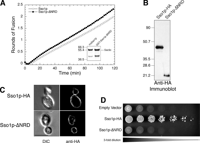
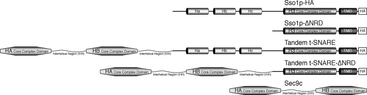
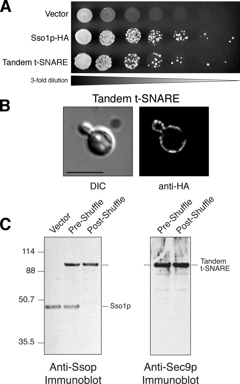
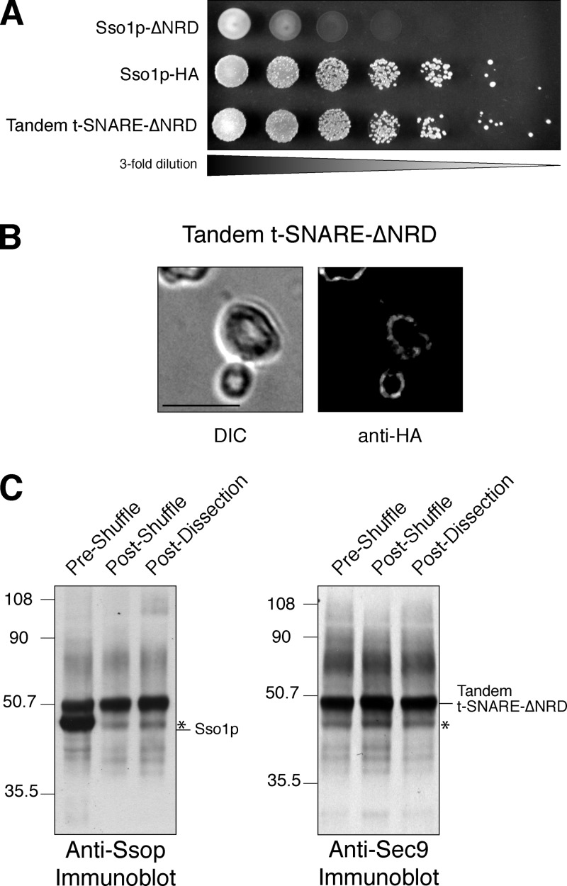
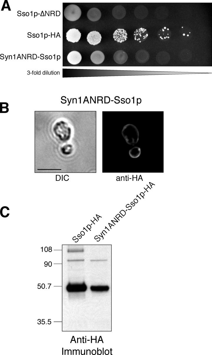
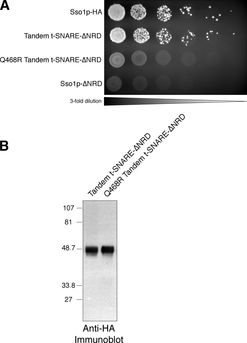
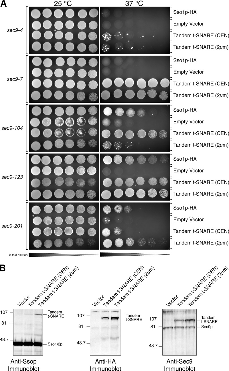
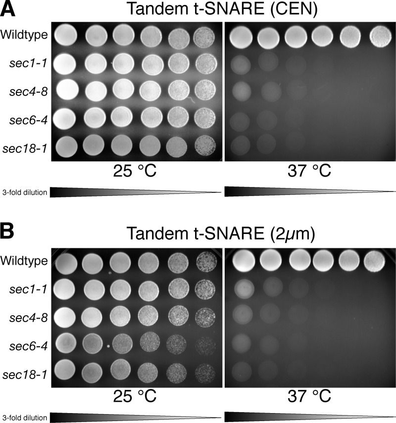
Membrane fusion in the secretory pathway is mediated by SNAREs (located on the vesicle membrane [v-SNARE] and the target membrane [t-SNARE]). In all cases examined, t-SNARE function is provided as a three-helix bundle complex containing three approximately 70-amino acid SNARE motifs. One SNARE motif is provided by a syntaxin family member (the t-SNARE heavy chain), and the other two helices are contributed by additional t-SNARE light chains. The syntaxin family is the most conformationally dynamic group of SNAREs and appears to be the major focus of SNARE regulation. An NH2-terminal region of plasma membrane syntaxins has been assigned as a negative regulatory element in vitro. This region is absolutely required for syntaxin function in vivo. We now show that the required function of the NH2-terminal regulatory domain (NRD) of the yeast plasma membrane syntaxin, Sso1p, can be circumvented when t-SNARE complex formation is made intramolecular. Our results suggest that the NRD is required for efficient t-SNARE complex formation and does not recruit necessary scaffolding factors.


Figures

Figure 1.
Figure 1.
Sso1p-ΔNRD is fusion competent in vitro but does not function in vivo. (A) In vitro fusion. Kinetic fusion graph comparing wild-type Sso1p (open circles) with Sso1p-ΔNRD (filled circles), illustrating that both are fusion competent with Snc1p fluorescent donor liposomes. (Inset) Coomassie-stained SDS-PAGE gel of the two t-SNARE liposomes. (B) Relative protein expression in yeast cell extracts of JMY303 and JMY305 was determined by immunoblot analysis detecting an HA epitope with the monoclonal antibody 16B12 (Covance). (C) Plasma membrane localization. Differential interference contrast (DIC) images and indirect immunofluorescence images are shown for JMY303 (Sso1p-HA) and JMY305 (Sso1p-ΔNRD). Localization was determined by staining with an anti-HA antibody. Bar, 5 μm. (D) Growth on 5-FOA. Three-fold serial dilutions of JMY302 (empty vector), JMY303 (Sso1p-HA), and JMY305 (Sso1pΔNRD) were spotted onto synthetic complete media with 2% galactose containing 1 mg/ml 5-FOA and grown at 30°C for 72 h.
Figure 2.
Figure 2.
Domain structure of t-SNARE proteins. Schematic representation of the domain structure of Sso1p, Sso1p-ΔNRD, Sec9c, and the chimeric tandem t-SNAREs. Sso1p-ΔNRD contains amino acids 179–290 of Sso1p. Sec9c is the SNAP25 homologous portion of Sec9p (amino acids 401–651). The tandem t-SNARE contains Sec9c and an additional copy of the Sec9p interhelical region (IHR; amino acids 499–588) covalently linked to the NH2 terminus of Sso1p. The additional Sec9p IHR allows sufficient conformational freedom to assume the necessary parallel orientation. The tandem t-SNARE–ΔNRD contains Sec9c, an additional copy of the Sec9p IHR (amino acids 499–588) linked to the NH2 terminus of Sso1p-ΔNRD. HA, helix A; HB, helix B; TMD, transmembrane domain.
Figure 3.
Figure 3.
The tandem t-SNARE can serve as the sole source of Ssop. (A) Growth on 5-FOA. Threefold serial dilutions of JMY302 (empty vector), JMY303 (Sso1p-HA), and JMY306 (tandem t-SNARE) were spotted onto synthetic complete media with 2% galactose containing 1 mg/ml 5-FOA and grown at 30°C for 72 h. (B) Plasma membrane localization. Differential interference contrast (DIC) images and indirect immunofluorescence images are shown for JMY306. Localization was determined by staining with an anti-HA antibody. Bar, 5 μm. (C) Whole cell extracts of JMY302 (empty vector), JMY306 (preshuffle), and JMY307 (postshuffle) were resolved by SDS-PAGE on a 4–10% BisTris NuPAGE gel and blotted with anti-Ssop antisera (∼29 μg of total protein per lane, left) or anti-Sec9p antisera (∼118 μg of total protein per lane, right). Note that endogenous Sec9p is not visualized because it is not efficiently transferred in this gel system.
Figure 4.
Figure 4.
The tandem t-SNARE–ΔNRD can function as the only source of Ssop. (A) Growth on 5-FOA. Threefold serial dilutions of JMY305 (Sso1 p-ΔNRD), JMY303 (Sso1p-HA), and JMY308 (tandem t-SNARE–ΔNRD) were spotted onto synthetic complete media with 2% galactose containing 1 mg/ml 5-FOA and grown at 30°C for 72 h. (B) Plasma membrane localization. Differential interference contrast (DIC) images and indirect immunofluorescence images are shown for JMY308. Localization was determined by staining with an anti-HA antibody. Bar, 5 μm. (C) Whole cell extracts of JMY308 (preshuffle), JMY309 (postshuffle), and JMY298 (postdissection) were resolved by SDS-PAGE on a 4–10% BisTris NuPAGE gel and blotted with anti-Ssop antisera (∼29 μg of total protein per lane, left) or anti-Sec9p antisera (∼115 μg of total protein per lane, right). The asterisk shows a tandem t-SNARE–ΔNRD proteolytic product that migrates at the same molecular weight as Ssop.
Figure 5.
Figure 5.
The syntaxin1A NRD cannot replace the Sso1p NRD in vivo. (A) Growth on 5-FOA. Threefold serial dilutions of JMY305 (Sso1p-ΔNRD), JMY303 (Sso1p-HA), and JMY310 (Syn1ANRD-Sso1p) were spotted onto synthetic complete media with 2% galactose containing 1 mg/ml 5-FOA and grown at 30°C for 72 h. (B) Plasma membrane localization. A differential interference contrast (DIC) image and an indirect immunofluorescence image are shown for JMY310. Localization was determined by staining with an anti-HA antibody. Bar, 5 μm. (C) Expression of the rSyn1A-Sso1p chimera. Whole cell extracts of JMY303 and JMY310 were prepared by glass bead lysis. Total protein (∼34 μg per lane) was resolved by SDS-PAGE on a 4–10% BisTris NuPAGE gel and probed with an anti-HA antibody.
Figure 6.
Figure 6.
The Q468R point mutation in the Sec9c segment of the tandem t-SNARE abolishes Sso1p function. (A) Growth on 5-FOA. Threefold serial dilutions of JMY303 (Sso1p-HA), JMY308 (tandem t-SNARE–ΔNRD), JMY334 (Q468R tandem t-SNARE–ΔNRD), and JMY305 (Sso1p-ΔNRD) were spotted onto synthetic complete media with 2% galactose containing 1 mg/ml 5-FOA and grown at 30°C for 72 h. (B) Whole cell extracts of JMY308 (tandem t-SNARE–ΔNRD) and JMY334 (Q468R tandem t-SNARE–ΔNRD) were resolved by SDS-PAGE on a 4–10% BisTris NuPAGE gel and blotted with an anti-HA antisera (∼21 μg of total protein per lane).
Figure 7.
Figure 7.
The tandem t-SNARE provides Sec9p function. (A) Threefold serial dilutions of each indicated cell culture (sec9-4 [JMY335-338], sec9-7 [JMY339-342], sec9-104 [JMY343-346], sec9-123 [JMY347-350], and sec9-201 [JMY351-354]) were spotted onto plates containing synthetic complete media with 2% glucose. One plate was grown at permissive temperature (25°C), and one was grown at restrictive temperature (37°C) for ∼2.5 d. The tandem t-SNARE efficiently complements the sec9-7ts, sec9-104ts, sec9-123ts, and sec9-201ts alleles but minimally complements the sec9-4ts allele. (B) Tandem t-SNARE expression levels. Whole cell extracts of JMY363 (vector), JMY364 (tandem t-SNARE; CEN), and JMY365 (tandem t-SNARE; 2 μm) were prepared by glass bead lysis. The indicated whole cell extracts were resolved by SDS-PAGE on a 4–10% BisTris NuPAGE gel (left and middle) and blotted with anti-Ssop antisera (∼95 μg of total protein per lane, left) or anti-HA antisera (∼75 μg of total protein per lane, middle). Additionally, a 7% standard gel (∼110 μg of total protein per lane, right) was probed with anti-Sec9 antisera.
Figure 8.
Figure 8.
The tandem t-SNARE does not suppress other late-acting sec mutants. Threefold serial dilutions of each indicated cell culture were spotted onto plates containing synthetic complete media with 2% glucose. Three late-acting sec mutants were transformed with the tandem t-SNARE in low copy (A; sec1-1 [JMY355], sec4-8 [JMY356], and sec6-4 [JMY357]) or high copy (B; sec1-1 [JMY359], sec4-8 [JMY360], and sec6-4 [JMY361]) and grown at permissive (25°C) or restrictive temperature (37°C) for ∼2.5 d. The general SNARE chaperone Sec18 (sec18-1 [JMY358 and JMY362]) was used as a control. No suppression was observed for these mutants.

Similar articles

Cited by

References

    1. Aalto, M.K., H. Ronne, and S. Keranen. 1993. Yeast syntaxins Sso1p and Sso2p belong to a family of related membrane proteins that function in vesicular transport. EMBO J. 12:4095–4104. - PMC - PubMed
    1. Bennett, W.F., N.F. Paoni, B.A. Keyt, D. Botstein, A.J. Jones, L. Presta, F.M. Wurm, and M.J. Zoller. 1991. High resolution analysis of functional determinants on human tissue-type plasminogen activator. J. Biol. Chem. 266:5191–5201. - PubMed
    1. Burke, D., D. Dawson, and T. Stearns. 2000. Methods in Yeast Genetics: a Cold Spring Harbor Laboratory Course Manual. Cold Spring Harbor Laboratory Press, Cold Spring Harbor, NY. xvii, 205 pp.
    1. Calakos, N., M.K. Bennett, K.E. Peterson, and R.H. Scheller. 1994. Protein-protein interactions contributing to the specificity of intracellular vesicular trafficking. Science. 263:1146–1149. - PubMed
    1. Chen, Y.A., and R.H. Scheller. 2001. SNARE-mediated membrane fusion. Nat. Rev. Mol. Cell Biol. 2:98–106. - PubMed

Publication types

MeSH terms