. 2006 Jan;147 Suppl 1(Suppl 1):S172-81.
doi: 10.1038/sj.bjp.0706429.

Purinergic signalling

Affiliations

Purinergic signalling

Geoffrey Burnstock. Br J Pharmacol. 2006 Jan.

Abstract

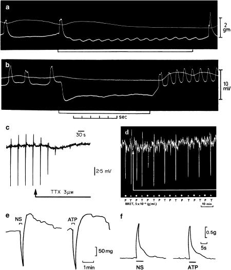
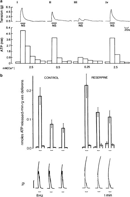
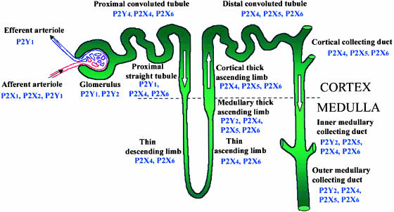
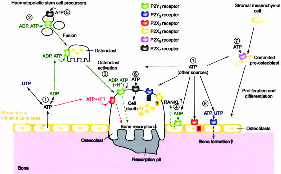
While there were early papers about the extracellular actions of purines, the role of ATP as a purinergic neurotransmitter in nonadrenergic, noncholinergic nerves in the gut and bladder in 1972 was a landmark discovery, although it met considerable resistance for the next 20 years. In the early 1990s, receptors for purines were cloned: four P1 receptor subtypes and seven P2X ionotropic and eight P2Y metabotropic receptor subtypes are currently recognized and characterized. The mechanisms underlying ATP release and breakdown are discussed. Purines and pyrimidines have major roles in the activities of non-neuronal cells as well as neurons. This includes fast signalling roles in exocrine and endocrine secretion, platelet aggregation, vascular endothelial cell-mediated vasodilation and nociceptive mechanosensory transduction, as well as acting as a cotransmitter and neuromodulator in most, if not all, nerve types in the peripheral and central nervous systems. More recently, slow (trophic) purinergic signalling has been implicated in cell proliferation, migration, differentiation and death in embryological development, wound healing, restenosis, atherosclerosis, ischaemia, cell turnover of epithelial cells in skin and visceral organs, inflammation, neuroprotection and cancer.


Figures

Figure 1
Figure 1
(a) and (b) Sucrose gap records from smooth muscle of guinea-pig taenia coli showing inhibitory junction potentials in response to stimulation of intrinsic nerves. Frequencies of stimulation for (a) 1/s and (b) 30/s. Upper trace tension, and lower trace membrane potential. Note the phase of excitation in (b) which follows cessation of stimulation (From Burnstock et al. (1963). Reproduced with kind permission from Nature Publishing Group). (c) Sucrose gap recording of membrane potential changes in smooth muscle of guinea-pig taenia coli in the presence of atropine (0.3 μM) and guanethidine (4 μM). Transmural field stimulation (0.5 ms, 0.033 Hz, 8 V) evoked transient hyperpolarisations or inhibitory junction potentials, which were followed by rebound depolarisations. Tetrodotoxin (TTX, 3 μM) added to the superfusing Krebs solution (applied at arrow) rapidly abolished the response to transmural field stimulation. (From Burnstock (1986). Acta Physiol. Scand. 126, 67–91. Reproduced with kind permission from Blackwell Publishing). (d) Effects of bretylium (BRET, 5 × 10⁻⁶ g ml−1) on responses to stimulation of the guinea-pig perivascular nerve–taenia coli preparation after atropine. Bretylium abolishes responses to stimulation of the perivascular nerves at 30 pulses/s (P, at dots), but only reduced responses to field stimulation of the taenia with 10 pulses/s (T, at triangles) (From Burnstock et al. (1966). Reproduced with kind permission from Blackwell Publishing). (e) Responses of the guinea-pig taenia coli to intramural nerve stimulation (NS, 1 Hz, 0.5 ms pulse duration, for 10 s at supramaximal voltage) and ATP (2 × 10⁻⁶M). The responses consist of a relaxation followed by a ‘rebound contraction'. Atropine (1.5 × 10⁻⁷M), guanethidine (5 × 10⁻⁶M) and sodium nitrite (7.2 × 10⁻⁴M) were present. (From Burnstock & Wong, 1978, Br. J. Pharmacol. 62, 293–302. Reproduced with kind permission from the Nature Publishing Group). (f) A comparison of the contractile responses of the guinea pig bladder strip to intramural nerve stimulation (NS: 5 Hz, 0.2 ms pulse duration and supramaximal voltage) and exogenous ATP (8.5 μM). Atropine (1.4 μM) and guanethidine (3.4 μM) were present throughout (From Burnstock et al., 1978. Reproduced with kind permission from the Nature Publishing Group).
Figure 2
Figure 2
(a) Effect of changing the calcium ion (Ca2+) concentration on the release of ATP from the guinea-pig isolated bladder strip during stimulation of intramural nerves. Upper trace: mechanical recording of changes in tension (g) during intramural nerve stimulation (NS: 20 Hz, 0.2 ms pulse duration, supramaximal voltage for 20 s). Lower trace: concentration of ATP in consecutive 20 s fractions of the superfusate. The Ca2+ concentration in the superfusate varied as follows: (i) 2.5 mM (normal Krebs); (ii) 0.5 mM; (iii) 0.25 mM; and (iv) 2.5 mM. The successive contractions were separated by 60 min intervals as indicated by the breaks in the mechanical trace. Atropine (1.4 μM) and guanethidine (3.4 μM) were present throughout. The temperature of the perfusate was between 22°C and 23°C (From Burnstock et al. (1978). Reproduced with kind permission from the Nature Publishing Group).(b) Release of endogenous ATP from control (n=32) and reserpine-treated (n=12) guinea-pig vasa deferentia during field stimulation at 8 Hz (pulse width 0.5 ms, 20 V). Upper panel: mean±s.e.m. nmol of ATP released min−1 g−1 of vas deferens. Lower panel: biphasic mechanical response to stimulation of the vas deferens for 1 min as denoted by the upward bracket. Note that the second slow phase of the mechanical response (mediated by NA) has gone in the reserpine-treated tissue (From Kirkpatrick & Burnstock (1987). Reproduced with kind permission from Elsevier).
Figure 3
Figure 3
Summary of the nephron segments immunopositive for P2 receptor subtypes (Modified from Turner, Vonend, Chan, Burnstock & Unwin, 2003. Cells, Tissues Organs 175, 105–117. Reproduced with kind permission from Karger AG, Basel).
Figure 4
Figure 4
Schematic diagram illustrating the potential roles played by extracellular nucleotides and P2 receptors in modulating bone cell function. Adenosine 5′-triphosphate (ATP), released from osteoblasts (e.g., through shear stress or constitutively) or from other sources, can be degraded to adenosine 5′-triphosphate (ADP) or converted into uridine 5′-triphosphate (UTP) via ecto-nucleotidases (1). All three nucleotides can act separately on specific P2 receptor subtypes, as indicated by the colour coding. ATP is a universal agonist, whereas UTP is only active at the P2Y2 receptor and ADP is only active at the P2Y1 receptor. ADP, via P2Y1 receptors, appears to stimulate both the formation (i.e., fusion) of osteoclasts from haematopoietic precursors (2) and the resorptive activity of mature osteoclasts (3). For the latter, a synergistic action of ATP and protons has also been proposed via the P2X2 receptor. ADP could also stimulate resorption indirectly through actions on osteoblasts, which in turn release pro-resorptive factors (e.g., receptor activator of nuclear factor kB ligand (RANKL)) (4). ATP at high concentrations might facilitate fusion of osteoclast progenitors through P2X7 receptor pore formation (5) or induce cell death of mature osteoclasts via P2X7 receptors (6). In osteoblasts, ATP, via P2X5 receptors, might enhance proliferation and/or differentiation (7). By contrast, UTP, via P2Y2 receptors, is a strong inhibitor of bone formation by osteoblasts (8). For some receptors (e.g., P2X4 and P2Y2 receptors on osteoclasts or P2X2 receptors on osteoblasts), evidence for expression has been found, but their role is still unclear (question marks). Dashed lines indicate signalling events in the cell. (From Hoebertz et al., 2003. Reproduced with kind permission from Elsevier).

Similar articles

Cited by

References

    1. ABBRACCHIO M.P., WILLIAMS M.(eds) (2001Handbook of Experimental Pharmacology: Purinergic and Pyrimidinergic Signalling, Vol. 151 (I and II) Berlin: Springer
    1. ABBRACCHIO M.P., BURNSTOCK G. Purinergic signalling: pathophysiological roles. Jpn. J. Pharmacol. 1998;78:113–145. - PubMed
    1. ABRAHAM E.H., SALIKHOVA A.Y., RAPAPORT E. ATP in the treatment of advanced cancer. Curr. Top. Membr. 2003;54:415–452.
    1. BAILEY M.A., TURNER C., HUS-CITHAREL A., MARCHETTI J., IMBERT-TEBOUL M., MILNER P., BURNSTOCK G., UNWIN R. P2Y receptors present in the native and isolated rat glomerulus. Nephron. Physiol. 2004;96:P79–P90. - PubMed
    1. BERNE R.M. Cardiac nucleotides in hypoxia: possible role in regulation of coronary blood flow. Am. J. Physiol. 1963;204:317–322. - PubMed

Publication types

MeSH terms