

. 2006 Jan;147 Suppl 1(Suppl 1):S27-37.
doi: 10.1038/sj.bjp.0706455.
G-protein-coupled receptors: past, present and future
Affiliations
- PMID: 16402114
- PMCID: PMC1760739
- DOI: 10.1038/sj.bjp.0706455
Item in Clipboard
G-protein-coupled receptors: past, present and future
Br J Pharmacol.
2006 Jan.
Abstract
The G-protein-coupled receptor (GPCR) family represents the largest and most versatile group of cell surface receptors. Drugs active at these receptors have therapeutic actions across a wide range of human diseases ranging from allergic rhinitis to pain, hypertension and schizophrenia. This review provides a brief historical overview of the properties and signalling characteristics of this important family of receptors.
Figures
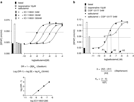
Examples of the use of the equations developed by Gaddum, Schild and Stephenson to determine the affinity constants of antagonists and partial agonists for GPCRs. (a) Antagonism by ICI 118551 of the CRE-reporter gene response (SPAP, secreted placental alkaline phosphatase) to salbutamol in CHO cells expressing the human β2-adrenoceptor. For each concentration of the antagonist ICI 118551, the ratio (DR, dose ratio) of salbutamol concentrations required to produce the same sized response (indicated by the grey line) in the presence and absence of the antagonist is determined. The antagonist affinity constant (KB) can then be determined either directly from the Gaddum equation or from a Schild plot of log (DR-1) against the log of the antagonist concentration ([B]). Data (unpublished observations) were kindly provided by Dr Jillian Baker. (b) Antagonism by CGP 12177 of the CRE-reporter gene response to salbutamol in CHO cells expressing the human β2-adrenoceptor. In this case, 3 nM CGP 12177 alone produces a partial agonist response. If the concentrations of salbutamol required to produce the same sized response in the presence (A3) and absence (A2) of CGP 12177 are determined along with the concentration of salbutamol alone that produces the same response as 3 nM CGP 12177 alone (A1), then the affinity constant of CGP 12177 (KP) can be determined as shown. In the original analysis by Stephenson (1956), he denoted x as the proportion of receptors occupied by partial agonist. The term shown as X therefore equates to (1−x) described in Stephenson's original formula. Data taken from Baker et al. (2002).
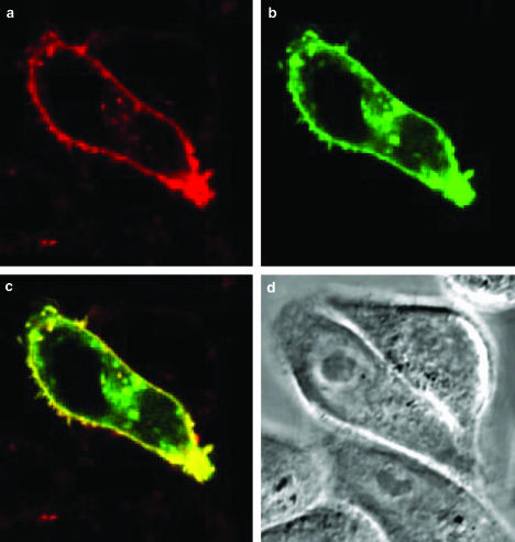
Binding of BODIPY-TMR-CGP12177 to CHO cells expressing the human β2-GFP adrenoceptor. (a) Binding of 50 nM BODIPY-TMR-CGP 12177 (red channel) to the same cells as in (b) which shows the location of the GFP-tagged β2-adrenoceptor (green channel). (c) An overlay of the two images (a, b) with colocalised pixels shown in yellow. (d) Phase contrast image of these cells (Baker J.G. & Hill, S.J. unpublished observations). The image was used as the poster for the British Pharmacological Society's 1st James Black Conference ‘Activation of cell surface receptors: new insights into ligand-gated ion channels and G-protein-coupled receptors', Cambridge, 2002.
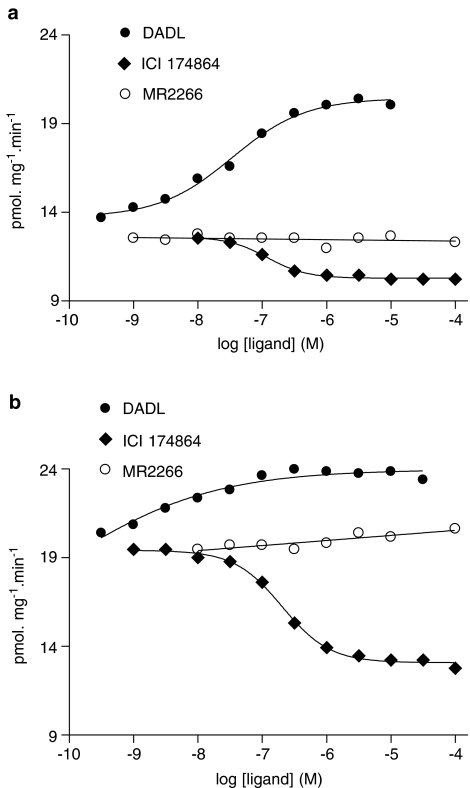
Agonist and inverse agonist effects of opioid ligands on GTPase activity in membranes of NG108-15 cells. High-affinity GTPase activity was measured in medium containing (a) 150 mM NaCl or (b) KCl. Data were taken from Costa & Herz, (1989). In the absence of NaCl (b), there is increased constitutive activity and the inverse agonist effects of ICI 174864 are more noticeable. MR 2266 was also able to competitively antagonise both the agonist effects of DADL and the inverse agonist effects of ICI 174864 (Costa & Herz, 1989). It therefore behaves as a neutral antagonist.
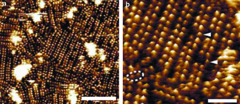
Organisation and topography of the cytoplasmic surface of rhodopsin. (a) Topograph obtained using atomic-force microscopy showing paracrystalline arrangement of rhodopsin dimers in a native disk membrane. (b) Magnification of a region of the topograph shown in (a) showing rows of rhodopsin dimers. Individual dimers (dashed elipse) and monomers (arrow heads) can also be observed. Scale bars (a) 50 nm and (b) 15 nm. Reproduced with permission from Fotiadis et al. (2003).
References
-
- BAKER J.G., HALL I.P., HILL S.J. Agonist and inverse agonist actions of β-blockers at the human β2-adrenoceptor provide evidence for agonist-directed signalling. Mol. Pharmacol. 2003;64:1357–1369. - PubMed
-
- BANERES J.L., PARELLO J. Structure-based analysis of GPCR function: evidence for a novel pentameric assembly between dimeric leukotriene B4 receptor BLT1 and the G-protein. J. Med. Biol. 2003;329:815–829. - PubMed
Publication types
MeSH terms
Substances
Grants and funding
LinkOut - more resources
Full Text Sources
Other Literature Sources
Miscellaneous
