. 2006 Jan 10:3:1.
doi: 10.1186/1742-4682-3-1.

Common angiotensin receptor blockers may directly modulate the immune system via VDR, PPAR and CCR2b

Affiliations

Common angiotensin receptor blockers may directly modulate the immune system via VDR, PPAR and CCR2b

Trevor G Marshall et al. Theor Biol Med Model. .

Abstract

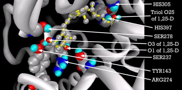
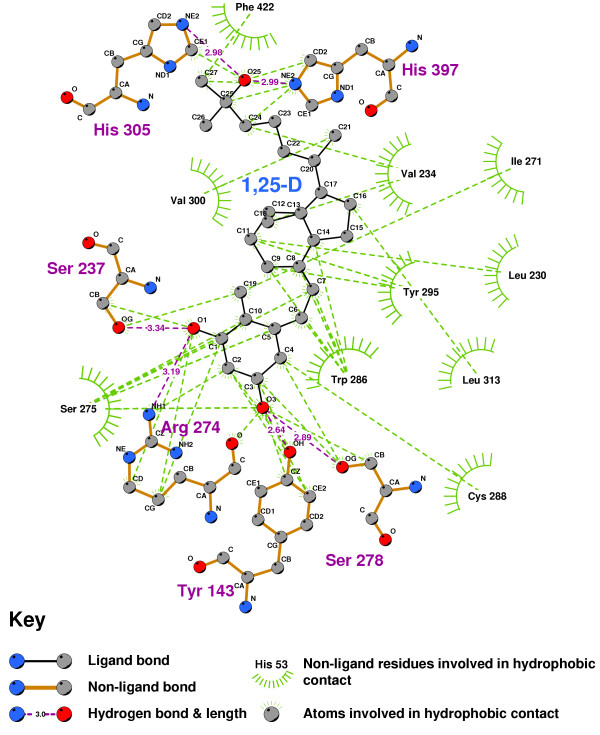
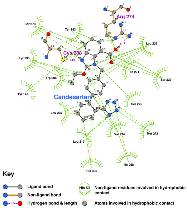
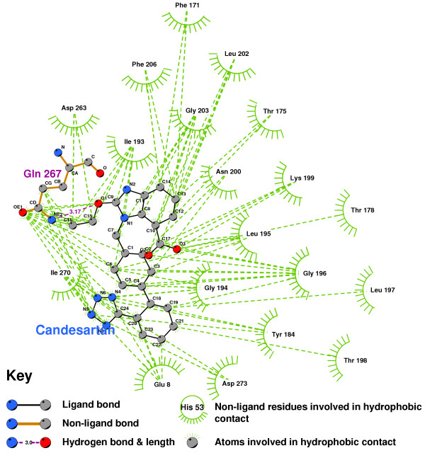
Background: There have been indications that common Angiotensin Receptor Blockers (ARBs) may be exerting anti-inflammatory actions by directly modulating the immune system. We decided to use molecular modelling to rapidly assess which of the potential targets might justify the expense of detailed laboratory validation. We first studied the VDR nuclear receptor, which is activated by the secosteroid hormone 1,25-dihydroxyvitamin-D. This receptor mediates the expression of regulators as ubiquitous as GnRH (Gonadatrophin hormone releasing hormone) and the Parathyroid Hormone (PTH). Additionally we examined Peroxisome Proliferator-Activated Receptor Gamma (PPARgamma), which affects the function of phagocytic cells, and the C-CChemokine Receptor, type 2b, (CCR2b), which recruits monocytes to the site of inflammatory immune challenge.

Results: Telmisartan was predicted to strongly antagonize (Ki asymptotically equal to 0.04 nmol) the VDR. The ARBs Olmesartan, Irbesartan and Valsartan (Ki asymptotically equal to10 nmol) are likely to be useful VDR antagonists at typical in-vivo concentrations. Candesartan (Ki asymptotically equal to 30 nmol) and Losartan (Ki asymptotically equal to 70 nmol) may also usefully inhibit the VDR. Telmisartan is a strong modulator of PPARgamma (Ki asymptotically equal to 0.3 nmol), while Losartan (Ki asymptotically equal to 3 nmol), Irbesartan (Ki asymptotically equal to 6 nmol), Olmesartan and Valsartan (Ki asymptotically equal to 12 nmol) also seem likely to have significant PPAR modulatory activity. Olmesartan and Irbesartan (Ki asymptotically equal to 9 nmol) additionally act as antagonists of a theoretical model of CCR2b. Initial validation of this CCR2b model was performed, and a proposed model for the Angiotensin II Type1 receptor (AT2R1) has been presented.

Conclusion: Molecular modeling has proven valuable to generate testable hypotheses concerning receptor/ligand binding and is an important tool in drug design. ARBs were designed to act as antagonists for AT2R1, and it was not surprising to discover their affinity for the structurally similar CCR2b. However, this study also found evidence that ARBs modulate the activation of two key nuclear receptors-VDR and PPARgamma. If our simulations are confirmed by experiment, it is possible that ARBs may become useful as potent anti-inflammatory agents, in addition to their current indication as cardiovascular drugs.


Figures

Figure 1
Figure 1
1,25-D and TX522 with superimposed X-ray and VDR-docked configurations. Note: Carbon atoms shown as grey, oxygen as red. Hydrogens not displayed.
Figure 2
Figure 2
VDR-docked configurations for 1,25-D and Telmisartan, separately and superimposed. Note: Models depicted as "thick" and "thin" solely for visual clarity. Carbon atoms shown as grey, oxygen as red, nitrogen shown as blue, polar hydrogen as blue-white. Non-polar hydrogens not displayed.
Figure 3
Figure 3
VDR-docked configurations for 1,25-D and Olmesartan, with superimposition showing both conformations. Note: Models depicted as "thick" and "thin" solely for visual clarity. Carbon atoms shown as grey, oxygen shown as red, nitrogen as blue, polar hydrogen as blue-white. Non-polar hydrogens not displayed.
Figure 4
Figure 4
VDR binding pocket showing primary 1,25-D docking residues. Note: 1,25-D depicted with yellow backbone for visual clarity. Carbon atoms shown as grey, oxygen as red, nitrogen as blue, polar hydrogen as blue-white. Non-polar hydrogens not displayed. Residues displayed as 'CPK' charge spheres, ligand in 'ball and stick' format.
Figure 5
Figure 5
2D LigPlot of 1,25-D bound into the VDR ligand binding pocket. Note: The core structure of the hydrogen-bonded residues is expanded to a 'ball-and-stick' format, so as to show the atoms involved in hydrogen bond formation.
Figure 6
Figure 6
The VDR agonist TX522 in the VDR ligand binding pocket. Note: The core structure of the hydrogen-bonded residues is expanded to a 'ball-and-stick' format, so as to show the atoms involved in hydrogen bond formation.
Figure 7
Figure 7
Olmesartan bound into the sterol terminus of the VDR binding pocket. Note: This is the 12 nanomolar conformation of Olmesartan in the binding pocket. The core structure of the hydrogen-bonded residues is expanded to a 'ball-and-stick' format, so as to show the atoms involved in hydrogen bond formation.
Figure 8
Figure 8
Telmisartan docked into the VDR ligand binding pocket. Note: Telmisartan is a strong antagonist of the VDR's activation.
Figure 9
Figure 9
Irbesartan docked into the VDR ligand binding pocket. Note: The core structure of the hydrogen-bonded residues is expanded to a 'ball-and-stick' format, so as to show the atoms involved in hydrogen bond formation.
Figure 11
Figure 11
Candesartan docked into the VDR ligand binding pocket. Note: The core structure of the hydrogen-bonded residues is expanded to a 'ball-and-stick' format, so as to show the atoms involved in hydrogen bond formation.
Figure 12
Figure 12
Losartan docked into the VDR ligand binding pocket. Note: The core structure of the hydrogen-bonded residues is expanded to a 'ball-and-stick' format, so as to show the atoms involved in hydrogen bond formation.
Figure 13
Figure 13
Overview of the ligand binding pocket identified in CCR2b (PDB:1KP1). Olmesartan is shown docked into pocket.
Figure 14
Figure 14
Perspective view showing how pocket is located underneath Extracellular 'loop' 1. Olmesartan is shown docked into pocket. Note: Residues displayed as 'CPK' charge spheres. Ligand displayed as stick and ball model. Left is view from front of pocket, facing helices 7 and 1, right view is from the top, looking across the top of helices 1 and 2.
Figure 15
Figure 15
CCR2b residues highlighted alongside docked TAK779. From left: front of pocket, rear of pocket. Note: Carbon atoms shown as grey, oxygen as red, nitrogen as blue, polar hydrogen as blue-white, sulphur as yellow. Non-polar hydrogens not displayed. Residues displayed as 'CPK' charge spheres, ligand as 'ball and stick' models.
Figure 16
Figure 16
TAK779 docked into the CCR2b binding pocket.
Figure 17
Figure 17
CCR2b residues highlighted alongside docked Olmesartan, viewed from the front of the binding pocket. Note: Carbon atoms shown as grey, oxygen as red, nitrogen as blue, polar hydrogen as blue-white, sulphur as yellow. Non-polar hydrogens not displayed. Residues displayed as 'CPK' charge spheres, ligand as 'ball and stick' models.
Figure 18
Figure 18
Olmesartan docked into the CCR2b binding pocket. Note: The core structure of the hydrogen-bonded residues is expanded to a 'ball-and-stick' format, so as to show the atoms involved in hydrogen bond formation.
Figure 19
Figure 19
CCR2b-docked configurations for TAK779 and Olmesartan, individually and with superimposition. Note: Ligands depicted as "thick" and "thin" solely for visual clarity. Carbon atoms shown as grey, oxygen as red, nitrogen as blue, polar hydrogen as blue-white. Non-polar hydrogens not displayed.
Figure 20
Figure 20
Irbesartan docked into the CCR2b binding pocket.
Figure 21
Figure 21
Putative AT2R1 with (from left) Olmesartan, and Losartan docked, showing primary residues. Ligands are also shown superimposed. Note: Carbon atoms shown as grey, oxygen as red, nitrogen as blue, polar hydrogen as blue-white, and chlorine as green. Non-polar hydrogens not displayed. Residues displayed as 'CPK' charge spheres, ligands as 'ball and stick' models. Thick and thin ligand backbones displayed solely for visual clarity.
Figure 22
Figure 22
Olmesartan docked into the putative AT2R1 binding pocket. Note: The core structure of the hydrogen-bonded residues is expanded to a 'ball-and-stick' format, so as to show the atoms involved in hydrogen bond formation.
Figure 23
Figure 23
Losartan docked into the putative AT2R1 binding pocket. Note: The core structure of the hydrogen-bonded residues is expanded to a 'ball-and-stick' format, so as to show the atoms involved in hydrogen bond formation.
Figure 24
Figure 24
Candesartan docked into the putative AT2R1 binding pocket. Note: The core structure of the hydrogen-bonded residues is expanded to a 'ball-and-stick' format, so as to show the atoms involved in hydrogen bond formation.
Figure 25
Figure 25
Farglitazar docked into the PPARgamma ligand binding pocket, showing the primary residues involved in hydrogenbonding. Note: Ligand depicted with yellow backbone solely for visual clarity. Carbon atoms shown as grey, oxygen as red, nitrogen as blue, polar hydrogen as blue-white. Non-polar hydrogens not displayed. Residues displayed as 'CPK' charge spheres, ligand as 'ball and stick' model.
Figure 26
Figure 26
Farglitazar docked into the PPARgamma ligand binding domain. Note: The core structure of the hydrogen-bonded residues is expanded to a 'ball-and-stick' format, so as to show the atoms involved in hydrogen bond formation.
Figure 27
Figure 27
Irbesartan docked into the PPARgamma ligand binding domain. Note: The core structure of the hydrogen-bonded residues is expanded to a 'ball-and-stick' format, so as to show the atoms involved in hydrogen bond formation.
Figure 28
Figure 28
Losartan docked into the PPARgamma ligand binding domain. Note: The core structure of the hydrogen-bonded residues is expanded to a 'ball-and-stick' format, so as to show the atoms involved in hydrogen bond formation.
Figure 29
Figure 29
Telmisartan docked into the PPARgamma ligand binding domain.
Figure 10
Figure 10
Valsartan docked into the VDR ligand binding pocket.

Similar articles

Cited by

References

    1. Mann DL, Deswal A. Angiotensin-receptor blockade in acute myocardial infarction – a matter of dose. N Engl J Med. 349:1963–5. doi: 10.1056/NEJMe038163. 2003 Nov 13. - DOI - PubMed
    1. United States Food and Drug Administration Approval Package for NDA21-286 http://www.fda.gov/cder/foi/nda/2002/21-286_Benicar_pharmr_P1.pdf Pharmacology Review, figures 1.1.1.1,1.1.1.3 and1.1.1.4.
    1. Izuhara Y, Nangaku M, Inagi R, Tominaga N, Aizawa T, Kurokawa K, van Ypersele de Strihou C, Miyata T. Renoprotective Properties of Angiotensin Receptor Blockers beyond Blood Pressure Lowering. J Am Soc Nephrol. 2005;16:3631–41. doi: 10.1681/ASN.2005050522. - DOI - PubMed
    1. Lewis EJ, Lewis JB. Treatment of diabetic nephropathy with angiotensin II receptor antagonist. Clin Exp Nephrol. 2003;7:1–8. doi: 10.1007/s101570300000. - DOI - PubMed
    1. Brenner BM, Cooper ME, de Zeeuw D, Keane WF, Mitch WE, Parving HH, et al. Effects of losartan on renal and cardiovascular outcomes in patients with type 2 diabetes and nephropathy. N Engl J Med. 2001;345:861–869. doi: 10.1056/NEJMoa011161. - DOI - PubMed

MeSH terms

LinkOut - more resources