. 2005;4(3):11.
doi: 10.1186/jbiol29. Epub 2005 Dec 14.

Compound developmental eye disorders following inactivation of TGFbeta signaling in neural-crest stem cells

Affiliations

Compound developmental eye disorders following inactivation of TGFbeta signaling in neural-crest stem cells

Lars M Ittner et al. J Biol. 2005.

Abstract

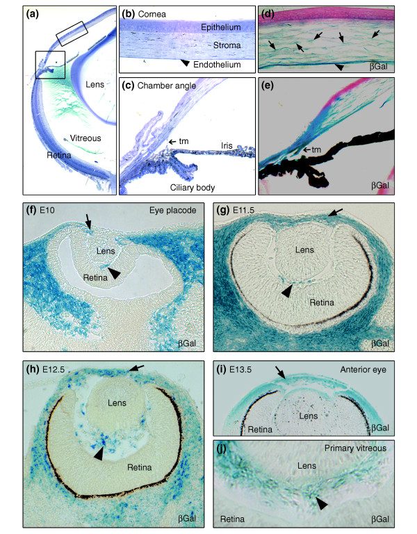
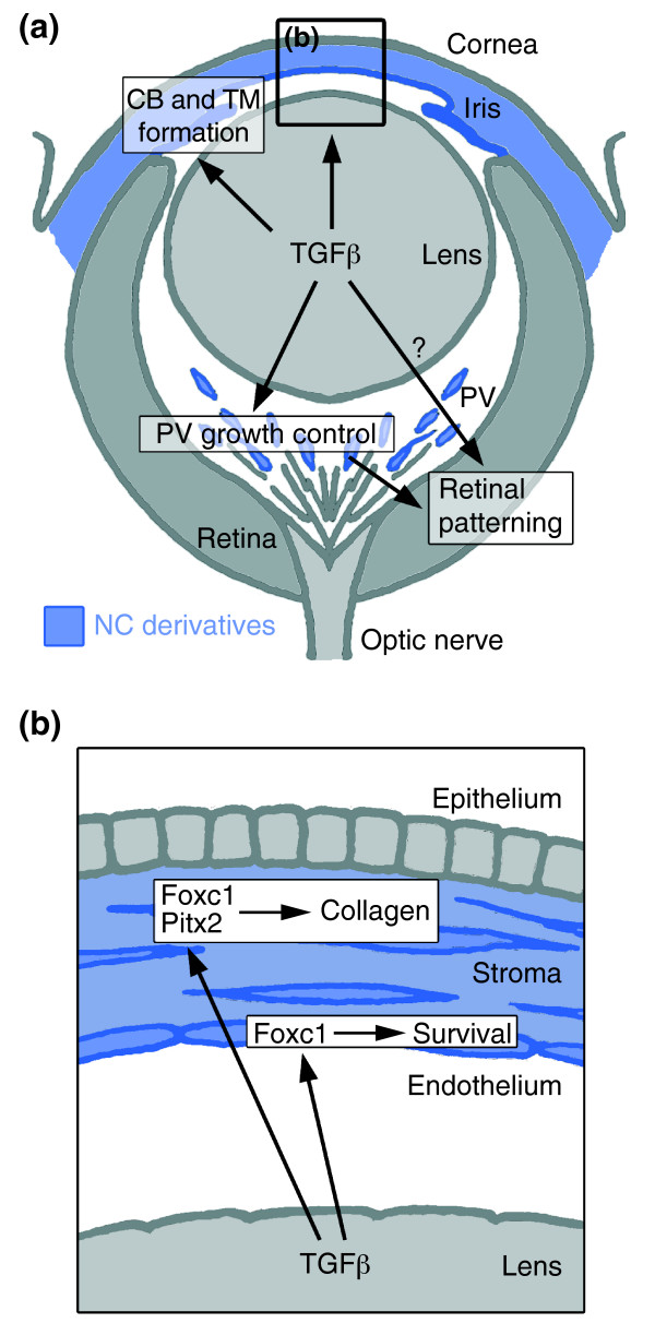
Background: Development of the eye depends partly on the periocular mesenchyme derived from the neural crest (NC), but the fate of NC cells in mammalian eye development and the signals coordinating the formation of ocular structures are poorly understood.

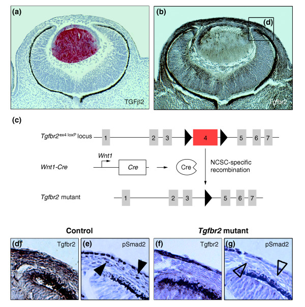
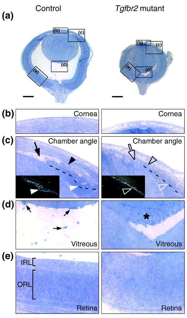
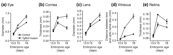
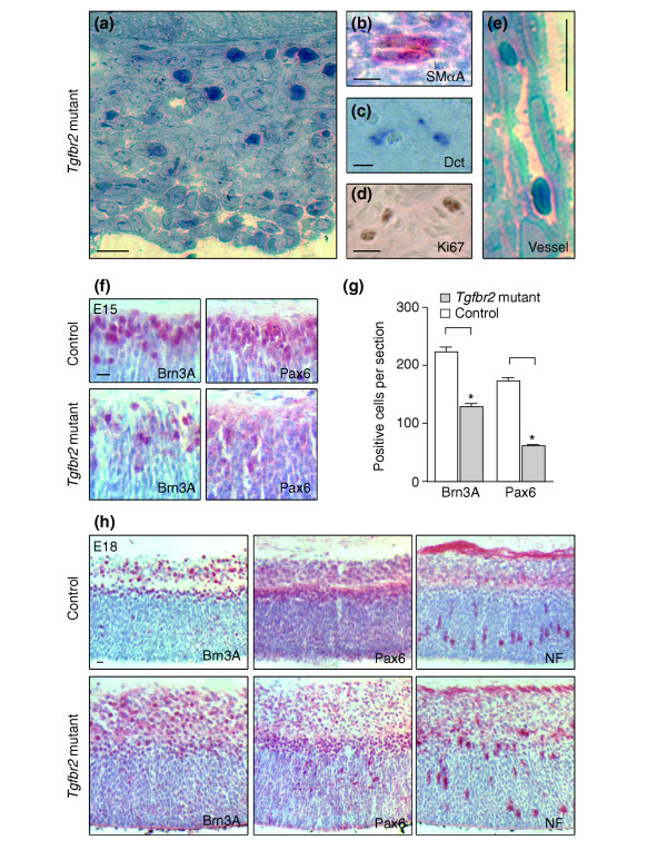
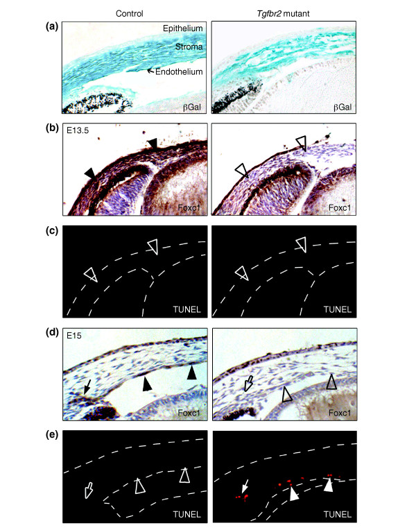
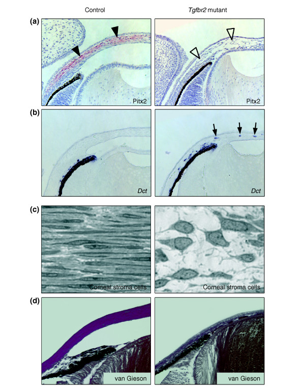
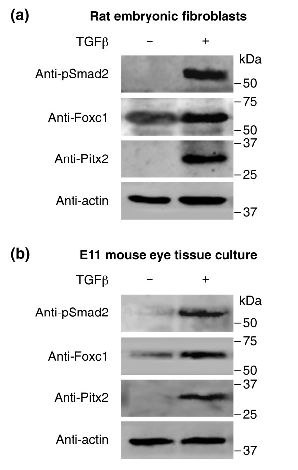
Results: Here we reveal distinct NC contributions to both anterior and posterior mesenchymal eye structures and show that TGFbeta signaling in these cells is crucial for normal eye development. In the anterior eye, TGFbeta2 released from the lens is required for the expression of transcription factors Pitx2 and Foxc1 in the NC-derived cornea and in the chamber-angle structures of the eye that control intraocular pressure. TGFbeta enhances Foxc1 and induces Pitx2 expression in cell cultures. As in patients carrying mutations in PITX2 and FOXC1, TGFbeta signal inactivation in NC cells leads to ocular defects characteristic of the human disorder Axenfeld-Rieger's anomaly. In the posterior eye, NC cell-specific inactivation of TGFbeta signaling results in a condition reminiscent of the human disorder persistent hyperplastic primary vitreous. As a secondary effect, retinal patterning is also disturbed in mutant mice.

Conclusion: In the developing eye the lens acts as a TGFbeta signaling center that controls the development of eye structures derived from the NC. Defective TGFbeta signal transduction interferes with NC-cell differentiation and survival anterior to the lens and with normal tissue morphogenesis and patterning posterior to the lens. The similarity to developmental eye disorders in humans suggests that defective TGFbeta signal modulation in ocular NC derivatives contributes to the pathophysiology of these diseases.


Figures

Figure 1
Figure 1
Neural crest (NC)-derived cells contribute to ocular development. (a) Toluidine blue staining of an adult eye. The boxed areas correspond to (b) a detailed view of the corneal assembly, including outer epithelium, stroma, and inner endothelium, and (c) the chamber angle at the irido-corneal transition which includes the trabecular meshwork (tm). (d-j) In vivo fate mapping of NC-derived, β-galactosidase (βGal)-expressing cells (blue) reveals (d) the NC origin of corneal keratocytes (arrows) and of corneal endothelium (arrowhead). (e) Structures of the chamber angle, including the trabecular meshwork are seen to be NC-derived. (f) At E10, the optic cup is surrounded by NC-derived cells expressing βGal. (g-i) The majority of the cells in the periocular mesenchyme (arrows), which forms the anterior eye segment, are of NC origin, as assessed from E11.5 to E13.5. (j) The primary vitreous at E13.5 (arrowheads) shows a strong NC contribution.
Figure 2
Figure 2
Inactivation of TGFβ signaling in ocular NC-derived cells. (a,b) TGFβ ligand and receptor expression in the developing eye at E13.5. (a) Immunoreactive TGFβ2 (red) is predominantly expressed in the lens, whereas (b) Tgfbr2 immunostaining (brown) shows a broad expression of the receptor in the forming eye, including the periocular mesenchyme, lens, primary vitreous, and retina. (c) Strategy used for Cre/loxP-mediated deletion of exon 4 of the Tgfbr2 locus in NC stem cells (NCSC). Exon 4 (red), encoding the transmembrane domain and the intracellular phosphorylation sites of the Tgfbr2 protein, is flanked by loxP sites (triangles) and deleted in NCSCs upon breeding with Wnt1-Cre mice. (d-g) A detailed view of the forming anterior eye segment (box in b). (d) Strong expression of Tgfbr2 (brown) in the prospective chamber angle, corneal stroma and endothelium can be seen in control embryos. (f) After deletion of Tgfbr2 in NCSC, Tgfbr2 is undetectable in corresponding structures. Moreover, defective TGFβ signaling in these structures is also reflected by the absence of phosphorylated pSmad2 in (g) Tgfbr2 mutant (open arrowheads) as compared with (e) control embryos (arrowheads).
Figure 3
Figure 3
Compound ocular anomalies in Tgfbr2-mutant mice. (a) Toluidine blue staining of semi-thin sagittal sections of eyes at E18 reveals a smaller size with no anterior chamber and an infiltration of cells behind the lens in Tgfbr2-mutant embryos as compared with control embryos. Boxes indicate magnified regions shown in the other panels; scale bars represent 250 μm. (b) Abnormal corneal stroma in Tgfbr2-mutant embryos. (c) Structures of the forming chamber angle, including the trabecular meshwork (black arrowhead) in control eyes are absent in Tgfbr2-mutant eyes (open black arrowhead). Here, the lens and the cornea fail to separate to form the anterior eye chamber (open arrow). In addition, dark-field images (insets) visualizing the pigment of the forming iris (broken line in the main image) reveal initiation of ciliary-body formation (white arrowheads) in control eyes and its absence in Tgfbr2-mutant eyes (open white arrowheads). (d) In control eyes, the primary vitreous consists of loosely arranged vessels of the hyaloid vascular system (arrows). In contrast, Tgfbr2-mutant mice show a dense cell mass between the lens and the retina (asterisk), reminiscent of human persistent hyperplastic primary vitreous. (e) The retina of control eyes displays typical patterning, with clear separation into an inner layer (IRL) and an outer layer (ORL). In Tgfbr2-mutant mice, however, there is no apparent patterning of the retina.
Figure 4
Figure 4
Impaired ocular growth in Tgfbr2-mutant mice leads to microphthalmia. (a) The developing eyes and (b-e) eye compartments of Tgfbr2-mutant and control embryos are of comparable size at E13.5, but subsequently, the eyes of Tgfbr2-mutant mice are smaller than controls. (c) The growth of the lens is comparable, but (b) the thickness of the cornea and (d) vitreous (measured as the distance between the lens and the optic-nerve disc) are drastically decreased in Tgfbr2-mutant mice. (e) In contrast, the thickness of the retina is increased in the mutant. For each time point, mid-organ sagittal sections of both eyes were analyzed for at least three mice.
Figure 5
Figure 5
Persistent hypertrophic primary vitreous and disturbed retinal patterning in Tgfbr2-mutant mice. (a) Detailed view of the persistent hypertrophic primary vitreous in E18 Tgfbr2-mutant mice, showing a dense retrolental cell mass. (b-d) Staining shows that this mass is composed of various cell types, including (b) smooth muscle α-actin (SMαA)-positive pericytes (red) and (c) prospective melanocytes expressing Dct mRNA (blue). (d) Ki67 staining indicates cell proliferation (brown). (e) The persistent hypertrophic primary vitreous contains vessels of the hyaloid vascular system. (f) Expression of Brn3A and Pax6 (red antibody staining) is readily detectable at E15 in the inner retinal layers of control eyes (top). In Tgfbr2-mutant eyes, however, cells expressing these markers are less frequent. (g) Bar graph of the results shown in (f). Asterisks indicate a significant difference (p < 0.001). (h) At E18, staining for Brn3A, Pax6, and neurofilaments (NF) reveals the expected patterning of the retina in control eyes and a diffuse distribution in Tgfbr2-mutant embryos. Thus, retinal patterning is disturbed in Tgfbr2-mutant embryos with persistent hypertrophic primary vitreous. Scale bars represent 10 μm.
Figure 6
Figure 6
Tgfbr2-mutant mice lack corneal expression of the transcription factor Foxc1. (a) In vivo fate mapping at E15 (βGal, blue) demonstrates that NC-derived cells have correctly migrated into control and Tgfbr2-mutant eyes, contributing to corneal stroma and endothelium. (b) At E13.5, the periocular mesenchyme of control eyes is positive for Foxc1 antibody staining (brown; arrowheads), whereas Foxc1 is undetectable in corresponding structures of Tgfbr2-mutant eyes (open arrowheads). (c) No apoptotic cells are found in either control or Tgfbr2-mutant eyes at E13.5 by TUNEL analysis (open arrowheads). (d) At E15, the eyes of control embryos show strong expression of Foxc1 (brown) in the forming trabecular meshwork (arrow) and in corneal endothelial cells (arrowheads). In Tgfbr2-mutant eyes, NC-derived cells localize to the cornea, but Foxc1 is undetectable in prospective endothelial cells (open arrowheads) and in the forming trabecular meshwork (open arrow). (e) At E15, cells that fail to express Foxc1 in Tgfbr2-mutant eyes appear to undergo apoptosis, unlike in control eyes, as revealed by TUNEL analysis (red).
Figure 7
Figure 7
Absence of the transcription factor Pitx2 and of collagen formation in the corneal stroma of Tgfbr2-mutant mice. (a) At E15, Pitx2 expression, as detected by immunohistochemistry (red; arrowheads) is restricted to corneal stromal cells in control animals but is undetectable in the corresponding structures of Tgfbr2-mutant mice (open arrowheads). (b) In situ hybridization for Dct, which marks prospective melanocytes, reveals atypical expression in the corneal stroma of Tgfbr2-mutant embryos (arrows). (c) High magnification of the corneal stroma shows the typical appearance of thin keratocytes in a parallel orientation and a dense extracellular matrix in control eyes at E18. In contrast, the corneal stroma of Tgfbr2-mutant embryos lacks extracellular matrix and has cells with large nuclei and a polygonal shape. (d) Van Gieson's staining reveals normal collagen matrix in the corneal stroma of control embryos (purple) that is absent in Tgfbr2-mutant embryos.
Figure 8
Figure 8
TGFβ regulates expression of Foxc1 and Pitx2. Western-blot analyses of cultured cells were performed using the antibodies shown. (a) Rat embryonic fibroblasts were treated with TGFβ, which results in increased levels of phosphorylated pSmad2. Furthermore, TGFβ signaling enhances expression of Foxc1 and induces Pitx2 expression, as revealed by western-blot analysis. (b) In ex vivo short-term tissue culture of E11 mouse eyes, including periocular mesenchyme, TGFβ strongly upregulates both Foxc1 and Pitx2 expression.
Figure 9
Figure 9
Summary of the TGFβ-dependent development of anterior and posterior ocular structures. (a) NC-derived cells (blue) contribute to structures of the anterior eye segment and the primary vitreous (PV). TGFβ signaling is involved in the formation of the ciliary body (CB) and the trabecular meshwork (TM), and in control of PV growth. Moreover, normal PV development and/or TGFβ signaling are important for correct retinal patterning. (b) In the cornea, prospective stromal keratocytes and endothelial cells are of NC origin. Here, TGFβ signaling is needed for the expression of the transcription factors Foxc1 and Pitx2 and for normal differentiation of NC-derived cells into collagen-synthesizing stromal keratocytes. Moreover, in forming corneal endothelial cells (and in the TM), expression of Foxc1 and cell survival requires TGFβ signaling.

Similar articles

Cited by

References

    1. Alward WL. Axenfeld-Rieger syndrome in the age of molecular genetics. Am J Ophthalmol. 2000;130:107–115. doi: 10.1016/S0002-9394(00)00525-0. - DOI - PubMed
    1. Kume T, Deng KY, Winfrey V, Gould DB, Walter MA, Hogan BL. The forkhead/winged helix gene Mf1 is disrupted in the pleiotropic mouse mutation congenital hydrocephalus. Cell. 1998;93:985–996. doi: 10.1016/S0092-8674(00)81204-0. - DOI - PubMed
    1. Lu MF, Pressman C, Dyer R, Johnson RL, Martin JF. Function of Rieger syndrome gene in left-right asymmetry and craniofacial development. Nature. 1999;401:276–278. doi: 10.1038/45797. - DOI - PubMed
    1. Holmberg J, Liu CY, Hjalt TA. PITX2 gain-of-function in Rieger syndrome eye model. Am J Pathol. 2004;165:1633–1641. - PMC - PubMed
    1. Tamimi Y, Lines M, Coca-Prados M, Walter MA. Identification of target genes regulated by FOXC1 using nickel agarose-based chromatin enrichment. Invest Ophthalmol Vis Sci. 2004;45:3904–3913. doi: 10.1167/iovs.04-0628. - DOI - PubMed

Publication types

MeSH terms