

Characterisation of the GRAF gene promoter and its methylation in patients with acute myeloid leukaemia and myelodysplastic syndrome
- PMID: 16404424
- PMCID: PMC2361128
- DOI: 10.1038/sj.bjc.6602939
Characterisation of the GRAF gene promoter and its methylation in patients with acute myeloid leukaemia and myelodysplastic syndrome
Abstract
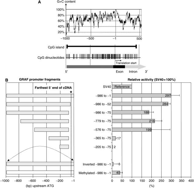
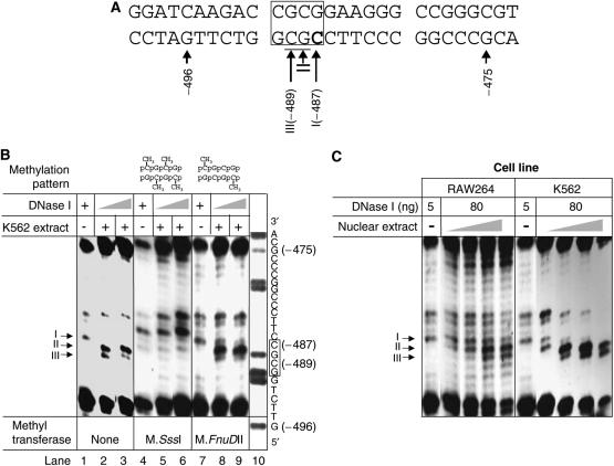
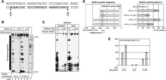
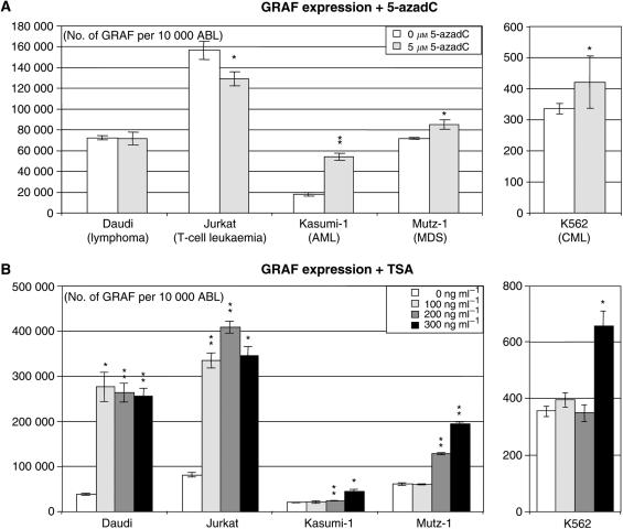
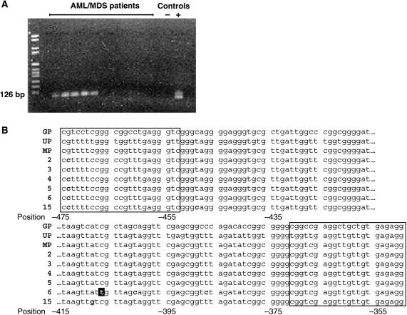
We report the isolation of the 5' flanking region of GRAF (GTPase regulator associated with the focal adhesion kinase), previously described as a putative tumour suppressor gene of acute myelogenous leukaemia and myelodysplastic syndrome, and demonstrate its promoter activity in reporter gene assays. Two putative protein-binding sites are identified of which one was sensitive to CpG methylation. The suppressed GRAF expression could be restored in leukaemia cell lines by treatment with a demethylating agent and an inhibitor of histone deacetylases. In contrast to normal tissues, which tested negative for GRAF promoter methylation, 11 of 29 (38%) bone marrow samples from patients with acute myeloid leukaemia or myelodysplastic syndrome were positive.
Figures
Similar articles
-
GTPase regulator associated with the focal adhesion kinase (GRAF) transcript was down-regulated in patients with myeloid malignancies.J Exp Clin Cancer Res. 2010 Aug 12;29(1):111. doi: 10.1186/1756-9966-29-111. J Exp Clin Cancer Res. 2010. PMID: 20704716 Free PMC article.
-
Abnormal methylation of GRAF promoter Chinese patients with acute myeloid leukemia.Leuk Res. 2011 Jun;35(6):783-6. doi: 10.1016/j.leukres.2010.10.013. Epub 2010 Nov 11. Leuk Res. 2011. PMID: 21074269
-
Aberrant methylation of GTPase regulator associated with the focal adhesion kinase (GRAF) promoter is an adverse prognostic factor in myelodysplastic syndrome.Eur J Haematol. 2010 Aug;85(2):174-6. doi: 10.1111/j.1600-0609.2010.01453.x. Epub 2010 Apr 1. Eur J Haematol. 2010. PMID: 20374274 No abstract available.
-
MicroRNAs in the pathogenesis of myelodysplastic syndromes and myeloid leukaemia.Curr Opin Hematol. 2014 Jul;21(4):276-82. doi: 10.1097/MOH.0000000000000054. Curr Opin Hematol. 2014. PMID: 24870972 Free PMC article. Review.
-
Treosulfan for acute myeloid leukaemia and myelodysplastic syndrome in adults, and malignant and non-malignant haematological diseases in children.Aust Prescr. 2023 Jun;46(1):18-19. doi: 10.18773/austprescr.2023.004. Aust Prescr. 2023. PMID: 38053669 Free PMC article. Review. No abstract available.
Cited by
-
Decreased expression of GRAF1/OPHN-1-L in the X-linked alpha thalassemia mental retardation syndrome.BMC Med Genomics. 2010 Jul 6;3:28. doi: 10.1186/1755-8794-3-28. BMC Med Genomics. 2010. PMID: 20602808 Free PMC article.
-
The oncocytic subtype is genetically distinct from other pancreatic intraductal papillary mucinous neoplasm subtypes.Mod Pathol. 2016 Sep;29(9):1058-69. doi: 10.1038/modpathol.2016.98. Epub 2016 Jun 10. Mod Pathol. 2016. PMID: 27282351 Free PMC article.
-
Fixing the GAP: The role of RhoGAPs in cancer.Eur J Cell Biol. 2022 Apr;101(2):151209. doi: 10.1016/j.ejcb.2022.151209. Epub 2022 Feb 10. Eur J Cell Biol. 2022. PMID: 35180567 Free PMC article. Review.
-
ADAR1 regulates ARHGAP26 gene expression through RNA editing by disrupting miR-30b-3p and miR-573 binding.RNA. 2013 Nov;19(11):1525-36. doi: 10.1261/rna.041533.113. Epub 2013 Sep 25. RNA. 2013. PMID: 24067935 Free PMC article.
-
GRAF1 deficiency blunts sarcolemmal injury repair and exacerbates cardiac and skeletal muscle pathology in dystrophin-deficient mice.Skelet Muscle. 2015 Aug 21;5:27. doi: 10.1186/s13395-015-0054-6. eCollection 2015. Skelet Muscle. 2015. PMID: 26301073 Free PMC article.
References
-
- Ammerpohl O, Short ML, Asbrand C, Schmitz A, Renkawitz R (1997) Complex protein binding to the mouse M-lysozyme gene downstream enhancer involves single-stranded DNA binding. Gene 200: 75–84 - PubMed
-
- Baylin SB, Herman JG (2000) DNA hypermethylation in tumorigenesis: epigenetics joins genetics. Trends Genet 16: 168–174 - PubMed
-
- Bednarik DP, Duckett C, Kim SU, Perez VL, Griffis K, Guenthner PC, Folks TM (1991) DNA CpG methylation inhibits binding of NF-kappa B proteins to the HIV- 1 long terminal repeat cognate DNA motifs. New Biol 3: 969–976 - PubMed
Publication types
MeSH terms
Substances
LinkOut - more resources
Full Text Sources
Medical
