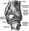
The anatomy and mechanisms of syndesmotic ankle sprains
- PMID: 16404437
- PMCID: PMC155405
The anatomy and mechanisms of syndesmotic ankle sprains
Abstract
Objective: To present a comprehensive review of the anatomy, biomechanics, and mechanisms of tibiofibular syndesmosis ankle sprains.
Data sources: MEDLINE (1966-1998) and CINAHL (1982-1998) searches using the key words syndesmosis, tibiofibular, ankle injuries, and ankle injuries-etiology.
Data synthesis: Stability of the distal tibiofibular syndesmosis is necessary for proper functioning of the ankle and lower extremity. Much of the ankle's stability is provided by the mortise formed around the talus by the tibia and fibula. The anterior and posterior inferior tibiofibular ligaments, the interosseous ligament, and the interosseous membrane act to statically stabilize the joint. During dorsiflexion, the wider portion anteriorly more completely fills the mortise, and contact between the articular surfaces is maximal. The distal structures of the lower leg primarily prevent lateral displacement of the fibula and talus and maintain a stable mortise. A variety of mechanisms individually or combined can cause syndesmosis injury. The most common mechanisms, individually and particularly in combination, are external rotation and hyperdorsiflexion. Both cause a widening of the mortise, resulting in disruption of the syndesmosis and talar instability. CONCLUSIONS AND RECOMMENDATION: Syndesmosis ankle injuries are less common than lateral ankle injuries, are difficult to evaluate, have a long recovery period, and may disrupt normal joint functioning. To effectively evaluate and treat this injury, clinicians should have a full understanding of the involved structures, functional anatomy, and etiologic factors.
Figures


References
-
- Singer KM, Jones DC, Taillon MR. Ligament injuries of the ankle and foot. In: Nicholas JA, Hershman EB, editors. The Lower Extremity and Spine in Sports Medicine. 2nd ed. Vol. 1. St Louis, MO: CV Mosby; 1995. pp. 423–440.
-
- Hopkinson WJ, St. Pierre P, Ryan JB, Wheeler JH. Syndesmosis sprains of the ankle. Foot Ankle. 1990;10:325–330. - PubMed
-
- Katznelson A, Lin E, Militiano J. Ruptures of the ligaments about the tibio-fibular syndesmosis. Injury. 1983;15:170–172. - PubMed
-
- Boytim MJ, Fischer DA, Neumann L. Syndesmotic ankle sprains. Am J Sports Med. 1991;19:294–298. - PubMed
-
- Taylor DC, Englehardt DL, Bassett FH., III Syndesmosis sprains of the ankle: the influence of heterotopic ossification. Am J Sports Med. 1992;20:146–150. - PubMed
LinkOut - more resources
Full Text Sources
